 Movie
Movie Controller

[English] 日本語
 Yorodumi
- PDB-6j8j: Structure of human voltage-gated sodium channel Nav1.7 in complex... -
+ Open data

- Basic information
| Entry | Database: PDB / ID: 6j8j | ||||||||||||||||||
|---|---|---|---|---|---|---|---|---|---|---|---|---|---|---|---|---|---|---|---|
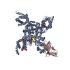
| Title | Structure of human voltage-gated sodium channel Nav1.7 in complex with auxiliary beta subunits, ProTx-II and tetrodotoxin (Y1755 down) | ||||||||||||||||||
 Components Components |
| ||||||||||||||||||
 Keywords Keywords |  MEMBRANE PROTEIN / voltage-gated sodium channel MEMBRANE PROTEIN / voltage-gated sodium channel | ||||||||||||||||||
| Function / homology |  Function and homology information Function and homology informationcorticospinal neuron axon guidance / positive regulation of voltage-gated sodium channel activity / response to pyrethroid / detection of mechanical stimulus involved in sensory perception / voltage-gated sodium channel activity involved in Purkinje myocyte action potential / regulation of sodium ion transmembrane transporter activity / voltage-gated sodium channel activity involved in cardiac muscle cell action potential / voltage-gated potassium channel activity involved in ventricular cardiac muscle cell action potential repolarization / membrane depolarization during Purkinje myocyte cell action potential / regulation of atrial cardiac muscle cell membrane depolarization ...corticospinal neuron axon guidance / positive regulation of voltage-gated sodium channel activity / response to pyrethroid / detection of mechanical stimulus involved in sensory perception / voltage-gated sodium channel activity involved in Purkinje myocyte action potential / regulation of sodium ion transmembrane transporter activity / voltage-gated sodium channel activity involved in cardiac muscle cell action potential / voltage-gated potassium channel activity involved in ventricular cardiac muscle cell action potential repolarization / membrane depolarization during Purkinje myocyte cell action potential / regulation of atrial cardiac muscle cell membrane depolarization / cardiac conduction / membrane depolarization during cardiac muscle cell action potential / positive regulation of sodium ion transport / node of Ranvier / membrane depolarization during action potential / cardiac muscle cell action potential involved in contraction / voltage-gated sodium channel complex / regulation of ventricular cardiac muscle cell membrane repolarization / locomotion / sodium channel inhibitor activity / neuronal action potential propagation / Interaction between L1 and Ankyrins / voltage-gated sodium channel activity / sodium ion transport / behavioral response to pain / Phase 0 - rapid depolarisation / regulation of heart rate by cardiac conduction / membrane depolarization / detection of temperature stimulus involved in sensory perception of pain / intercalated disc / sodium channel regulator activity / sodium ion transmembrane transport / neuronal action potential / cardiac muscle contraction / sensory perception of pain / T-tubule / post-embryonic development / axon guidance / response to toxic substance / Sensory perception of sweet, bitter, and umami (glutamate) taste / positive regulation of neuron projection development / circadian rhythm / nervous system development / gene expression / response to heat / chemical synaptic transmission / perikaryon / transmembrane transporter binding / cell adhesion / inflammatory response / axon / synapse / extracellular region / plasma membraneSimilarity search - Function | ||||||||||||||||||
| Biological species |  Homo sapiens (human) Homo sapiens (human) | ||||||||||||||||||
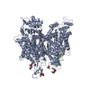
| Method | ELECTRON MICROSCOPY / single particle reconstruction / cryo EM / Resolution: 3.2 Å | ||||||||||||||||||
 Authors Authors | Shen, H. / Liu, D. / Lei, J. / Yan, N. | ||||||||||||||||||
| Funding support |  China, 5items China, 5items
| ||||||||||||||||||
 Citation Citation | Journal: Science / Year: 2019 Title: Structures of human Na1.7 channel in complex with auxiliary subunits and animal toxins. Authors: Huaizong Shen / Dongliang Liu / Kun Wu / Jianlin Lei / Nieng Yan / Abstract: Voltage-gated sodium channel Na1.7 represents a promising target for pain relief. Here we report the cryo-electron microscopy structures of the human Na1.7-β1-β2 complex bound to two combinations ...Voltage-gated sodium channel Na1.7 represents a promising target for pain relief. Here we report the cryo-electron microscopy structures of the human Na1.7-β1-β2 complex bound to two combinations of pore blockers and gating modifier toxins (GMTs), tetrodotoxin with protoxin-II and saxitoxin with huwentoxin-IV, both determined at overall resolutions of 3.2 angstroms. The two structures are nearly identical except for minor shifts of voltage-sensing domain II (VSD), whose S3-S4 linker accommodates the two GMTs in a similar manner. One additional protoxin-II sits on top of the S3-S4 linker in VSD The structures may represent an inactivated state with all four VSDs "up" and the intracellular gate closed. The structures illuminate the path toward mechanistic understanding of the function and disease of Na1.7 and establish the foundation for structure-aided development of analgesics. | ||||||||||||||||||
| History |
|
- Structure visualization
| Movie |
Movie viewer |
|---|---|
| Structure viewer | Molecule: MolmilJmol/JSmol |
- Downloads & links
-Download
| PDBx/mmCIF format | 6j8j.cif.gz | 320.4 KB | Display | PDBx/mmCIF format |
|---|---|---|---|---|
| PDB format | pdb6j8j.ent.gz | 243.7 KB | Display | PDB format |
| PDBx/mmJSON format | 6j8j.json.gz | Tree view | PDBx/mmJSON format | |
| Others |  Other downloads Other downloads |
-Validation report
| Arichive directory | https://data.pdbj.org/pub/pdb/validation_reports/j8/6j8jftp://data.pdbj.org/pub/pdb/validation_reports/j8/6j8j | HTTPS FTP |
|---|
-Related structure data
| Related structure data | 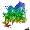 9782MC 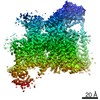 9781C  6j8gC  6j8hC  6j8iC M: map data used to model this data C: citing same article ( |
|---|---|
| Similar structure data |


-Links
 PDBj
PDBj


- Assembly
| Deposited unit | 
|
|---|---|
| 1 |
|
-Components
-Sodium channel subunit beta- ... , 2 types, 2 molecules CB
| #1: Protein | Mass: 24355.859 Da / Num. of mol.: 1 Source method: isolated from a genetically manipulated source Source: (gene. exp.) Homo sapiens (human) / Gene: SCN2B, UNQ326/PRO386 / Production host: Homo sapiens (human) / References: UniProt: O60939 |
|---|---|
| #3: Protein | Mass: 24732.115 Da / Num. of mol.: 1 Source method: isolated from a genetically manipulated source Source: (gene. exp.) Homo sapiens (human) / Gene: SCN1B / Production host: Homo sapiens (human) / References: UniProt: Q07699 |
-Protein / Non-polymers , 2 types, 2 molecules A
| #2: Protein | / Neuroendocrine sodium channel / hNE-Na / Peripheral sodium channel 1 / PN1 / Sodium channel protein ...Neuroendocrine sodium channel / hNE-Na / Peripheral sodium channel 1 / PN1 / Sodium channel protein type IX subunit alpha / Voltage-gated sodium channel subunit alpha Nav1.7 Mass: 231211.922 Da / Num. of mol.: 1 Source method: isolated from a genetically manipulated source Source: (gene. exp.) Homo sapiens (human) / Gene: SCN9A, NENA / Production host: Homo sapiens (human) / References: UniProt: Q15858 |
|---|---|
| #6: Chemical | ChemComp-9SR / (Tetrodotoxin Mass: 319.268 Da / Num. of mol.: 1 / Source method: obtained synthetically / Formula: C11H17N3O8 / Comment: neurotoxin*YM Mass: 319.268 Da / Num. of mol.: 1 / Source method: obtained synthetically / Formula: C11H17N3O8 / Comment: neurotoxin*YM |
-Sugars , 2 types, 9 molecules 
| #4: Polysaccharide | Source method: isolated from a genetically manipulated source #5: Sugar | ChemComp-NAG / N-Acetylglucosamine Type: D-saccharide, beta linking / Mass: 221.208 Da / Num. of mol.: 7 Type: D-saccharide, beta linking / Mass: 221.208 Da / Num. of mol.: 7Source method: isolated from a genetically manipulated source Formula: C8H15NO6 |
|---|
-Experimental details
-Experiment
| Experiment | Method: ELECTRON MICROSCOPY |
|---|---|
| EM experiment | Aggregation state: PARTICLE / 3D reconstruction method: single particle reconstruction |
- Sample preparation
| Component | Name: Complex of human voltage-gated sodium channel Nav1.7 with auxiliary beta subunits, ProTx-II and tetrodotoxin Type: COMPLEX / Entity ID: #1-#3 / Source: RECOMBINANT |
|---|---|
| Source (natural) | Organism: Homo sapiens (human) |
| Source (recombinant) | Organism: Homo sapiens (human) |
| Buffer solution | pH: 7.4 |
| Specimen | Embedding applied: NO / Shadowing applied: NO / Staining applied: NO / Vitrification applied: YES |
| Vitrification | Cryogen name: ETHANE |
- Electron microscopy imaging
| Experimental equipment |  Model: Titan Krios / Image courtesy: FEI Company |
|---|---|
| Microscopy | Model: FEI TITAN KRIOS |
| Electron gun | Electron source: FIELD EMISSION GUN / Accelerating voltage: 300 kV / Illumination mode: FLOOD BEAM |
| Electron lens | Mode: BRIGHT FIELDBright-field microscopy |
| Image recording | Electron dose: 48 e/Å2 / Film or detector model: GATAN K2 QUANTUM (4k x 4k) |
- Processing
| Software | Name: PHENIX / Version: 1.11.1_2575: / Classification: refinement | ||||||||||||||||||||||||
|---|---|---|---|---|---|---|---|---|---|---|---|---|---|---|---|---|---|---|---|---|---|---|---|---|---|
| CTF correction | Type: PHASE FLIPPING AND AMPLITUDE CORRECTION | ||||||||||||||||||||||||
| 3D reconstruction | Resolution: 3.2 Å / Resolution method: FSC 0.143 CUT-OFF / Num. of particles: 263205 / Symmetry type: POINT | ||||||||||||||||||||||||
| Refine LS restraints |
|
