 Movie
Movie Controller

[English] 日本語
 Yorodumi
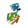
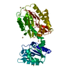
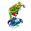
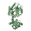
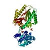
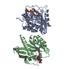
- PDB-6j0k: Crystal structure of intracellular B30.2 domain of BTN3A3 mutant ... -
+ Open data

- Basic information
| Entry | Database: PDB / ID: 6j0k | ||||||
|---|---|---|---|---|---|---|---|
| Title | Crystal structure of intracellular B30.2 domain of BTN3A3 mutant in complex with HMBPP | ||||||
 Components Components | Butyrophilin subfamily 3 member A3 | ||||||
 Keywords Keywords |  SIGNALING PROTEIN / Butyrophilin SIGNALING PROTEIN / Butyrophilin | ||||||
| Function / homology |  Function and homology information Function and homology informationButyrophilin (BTN) family interactions / T cell mediated immunity / regulation of cytokine production / T cell receptor signaling pathway / external side of plasma membrane / signaling receptor binding / membrane / plasma membraneSimilarity search - Function | ||||||
| Biological species |  Homo sapiens (human) Homo sapiens (human) | ||||||
| Method | X-RAY DIFFRACTION / SYNCHROTRON / MOLECULAR REPLACEMENT / Resolution: 2 Å | ||||||
 Authors Authors | Yang, Y.Y. / Liu, W.D. / Cai, N.N. / Chen, C.C. / Guo, R.T. / Zhang, Y.H. | ||||||
 Citation Citation | Journal: Immunity / Year: 2019 Title: A Structural Change in Butyrophilin upon Phosphoantigen Binding Underlies Phosphoantigen-Mediated V gamma 9V delta 2 T Cell Activation. Authors: Yang, Y. / Li, L. / Yuan, L. / Zhou, X. / Duan, J. / Xiao, H. / Cai, N. / Han, S. / Ma, X. / Liu, W. / Chen, C.C. / Wang, L. / Li, X. / Chen, J. / Kang, N. / Chen, J. / Shen, Z. / Malwal, S. ...Authors: Yang, Y. / Li, L. / Yuan, L. / Zhou, X. / Duan, J. / Xiao, H. / Cai, N. / Han, S. / Ma, X. / Liu, W. / Chen, C.C. / Wang, L. / Li, X. / Chen, J. / Kang, N. / Chen, J. / Shen, Z. / Malwal, S.R. / Liu, W. / Shi, Y. / Oldfield, E. / Guo, R.T. / Zhang, Y. | ||||||
| History |
|
- Structure visualization
| Structure viewer | Molecule: MolmilJmol/JSmol |
|---|
- Downloads & links
-Download
| PDBx/mmCIF format | 6j0k.cif.gz | 97.3 KB | Display | PDBx/mmCIF format |
|---|---|---|---|---|
| PDB format | pdb6j0k.ent.gz | 71.8 KB | Display | PDB format |
| PDBx/mmJSON format | 6j0k.json.gz | Tree view | PDBx/mmJSON format | |
| Others |  Other downloads Other downloads |
-Validation report
| Arichive directory | https://data.pdbj.org/pub/pdb/validation_reports/j0/6j0kftp://data.pdbj.org/pub/pdb/validation_reports/j0/6j0k | HTTPS FTP |
|---|
-Related structure data
| Related structure data |  5zxkC  5zz3C  6ismC 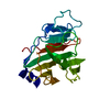 6itaC  6j06C 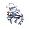 6j0gC  6j0lC  4n7uS S: Starting model for refinement C: citing same article ( |
|---|---|
| Similar structure data |


-Links
 PDBj
PDBj
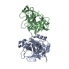
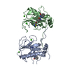
- Assembly
| Deposited unit | 
| ||||||||
|---|---|---|---|---|---|---|---|---|---|
| 1 |
| ||||||||
| 2 | 
| ||||||||
| Unit cell |
|
-Components
| #1: Protein | Mass: 25272.418 Da / Num. of mol.: 2 / Fragment: UNP residues 328-515 / Mutation: R351H Source method: isolated from a genetically manipulated source Source: (gene. exp.) Homo sapiens (human) / Gene: BTN3A3, BTF3 / Production host:  Escherichia coli BL21(DE3) (bacteria) / Strain (production host): BL21(DE3) / References: UniProt: O00478 Escherichia coli BL21(DE3) (bacteria) / Strain (production host): BL21(DE3) / References: UniProt: O00478#2: Chemical | (E)-4-Hydroxy-3-methyl-but-2-enyl pyrophosphate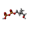  Mass: 262.092 Da / Num. of mol.: 2 / Source method: obtained synthetically / Formula: C5H12O8P2 Mass: 262.092 Da / Num. of mol.: 2 / Source method: obtained synthetically / Formula: C5H12O8P2#3: Water | ChemComp-HOH / | Water Mass: 18.015 Da / Num. of mol.: 182 / Source method: isolated from a natural source / Formula: H2O Mass: 18.015 Da / Num. of mol.: 182 / Source method: isolated from a natural source / Formula: H2OSequence details | The intracellular B30.2 domain of BTN3A3 was redesigned to have TEV protease cleavage sites and ...The intracellular B30.2 domain of BTN3A3 was redesigned to have TEV protease cleavage sites and bordering the linker region(AGAGA). | |
|---|
-Experimental details
-Experiment
| Experiment | Method: X-RAY DIFFRACTION / Number of used crystals: 1 |
|---|
- Sample preparation
| Crystal | Density Matthews: 1.76 Å3/Da / Density % sol: 30.22 % |
|---|---|
| Crystal grow | Temperature: 298 K / Method: vapor diffusion, sitting drop / Details: Isopropanol ,Imidazole, PEG 8000 |
-Data collection
| Diffraction | Mean temperature: 100 K / Serial crystal experiment: N |
|---|---|
| Diffraction source | Source: SYNCHROTRON / Site: NSRRC  / Beamline: BL15A1 / Wavelength: 1 Å / Beamline: BL15A1 / Wavelength: 1 Å |
| Detector | Type: RAYONIX MX-300 / Detector: CCD / Date: Nov 29, 2017 |
| Radiation | Protocol: SINGLE WAVELENGTH / Monochromatic (M) / Laue (L): M / Scattering type: x-ray |
| Radiation wavelength | Wavelength: 1 Å / Relative weight: 1 |
| Reflection | Resolution: 2→25 Å / Num. obs: 20422 / % possible obs: 84.2 % / Redundancy: 3 % / Rmerge(I) obs: 0.08 / Net I/σ(I): 16.06 |
| Reflection shell | Resolution: 2→2.07 Å / Num. unique obs: 2264 / CC1/2: 0.946 |
- Processing
| Software |
| ||||||||||||||||||||||||||||||||||||||||||||||||||||||||||||||||||||||||||||||||||||||||||||||||||||||||||||||||||||||||||||||||||||||||||||||||||||||||||||||||||||||||||||||||||||||
|---|---|---|---|---|---|---|---|---|---|---|---|---|---|---|---|---|---|---|---|---|---|---|---|---|---|---|---|---|---|---|---|---|---|---|---|---|---|---|---|---|---|---|---|---|---|---|---|---|---|---|---|---|---|---|---|---|---|---|---|---|---|---|---|---|---|---|---|---|---|---|---|---|---|---|---|---|---|---|---|---|---|---|---|---|---|---|---|---|---|---|---|---|---|---|---|---|---|---|---|---|---|---|---|---|---|---|---|---|---|---|---|---|---|---|---|---|---|---|---|---|---|---|---|---|---|---|---|---|---|---|---|---|---|---|---|---|---|---|---|---|---|---|---|---|---|---|---|---|---|---|---|---|---|---|---|---|---|---|---|---|---|---|---|---|---|---|---|---|---|---|---|---|---|---|---|---|---|---|---|---|---|---|---|
| Refinement | Method to determine structure: MOLECULAR REPLACEMENT Starting model: 4N7U Resolution: 2→25 Å / Cor.coef. Fo:Fc: 0.954 / Cor.coef. Fo:Fc free: 0.912 / SU B: 4.815 / SU ML: 0.139 / Cross valid method: THROUGHOUT / ESU R: 0.299 / ESU R Free: 0.226 / Details: HYDROGENS HAVE BEEN ADDED IN THE RIDING POSITIONS
| ||||||||||||||||||||||||||||||||||||||||||||||||||||||||||||||||||||||||||||||||||||||||||||||||||||||||||||||||||||||||||||||||||||||||||||||||||||||||||||||||||||||||||||||||||||||
| Solvent computation | Ion probe radii: 0.8 Å / Shrinkage radii: 0.8 Å / VDW probe radii: 1.2 Å | ||||||||||||||||||||||||||||||||||||||||||||||||||||||||||||||||||||||||||||||||||||||||||||||||||||||||||||||||||||||||||||||||||||||||||||||||||||||||||||||||||||||||||||||||||||||
| Displacement parameters | Biso mean: 25.298 Å2
| ||||||||||||||||||||||||||||||||||||||||||||||||||||||||||||||||||||||||||||||||||||||||||||||||||||||||||||||||||||||||||||||||||||||||||||||||||||||||||||||||||||||||||||||||||||||
| Refinement step | Cycle: 1 / Resolution: 2→25 Å
| ||||||||||||||||||||||||||||||||||||||||||||||||||||||||||||||||||||||||||||||||||||||||||||||||||||||||||||||||||||||||||||||||||||||||||||||||||||||||||||||||||||||||||||||||||||||
| Refine LS restraints |
|