 Movie
Movie Controller

[English] 日本語
 Yorodumi
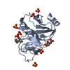
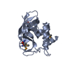
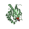
- PDB-4n7u: Crystal Structure of Intracellular B30.2 Domain of BTN3A1 in Comp... -
+ Open data

- Basic information
| Entry | Database: PDB / ID: 4n7u | ||||||
|---|---|---|---|---|---|---|---|
| Title | Crystal Structure of Intracellular B30.2 Domain of BTN3A1 in Complex with CHDMAPP | ||||||
 Components Components | Butyrophilin subfamily 3 member A1 | ||||||
 Keywords Keywords |  SIGNALING PROTEIN / Butyrophilin / CD277 / Phosphoantigen / B30.2 / PRY/SPRY SIGNALING PROTEIN / Butyrophilin / CD277 / Phosphoantigen / B30.2 / PRY/SPRY | ||||||
| Function / homology |  Function and homology information Function and homology informationButyrophilin (BTN) family interactions / activated T cell proliferation / regulation of cytokine production / positive regulation of cytokine production / positive regulation of type II interferon production / T cell receptor signaling pathway / adaptive immune response / external side of plasma membrane / signaling receptor binding / plasma membraneSimilarity search - Function | ||||||
| Biological species |  Homo sapiens (human) Homo sapiens (human) | ||||||
| Method | X-RAY DIFFRACTION / SYNCHROTRON / MOLECULAR REPLACEMENT / Resolution: 1.4598 Å | ||||||
 Authors Authors | Sandstrom, A. / Adams, E.J. | ||||||
 Citation Citation | Journal: Immunity / Year: 2014 Title: The Intracellular B30.2 Domain of Butyrophilin 3A1 Binds Phosphoantigens to Mediate Activation of Human V gamma 9V delta 2 T Cells. Authors: Sandstrom, A. / Peigne, C.M. / Leger, A. / Crooks, J.E. / Konczak, F. / Gesnel, M.C. / Breathnach, R. / Bonneville, M. / Scotet, E. / Adams, E.J. | ||||||
| History |
|
- Structure visualization
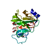
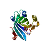
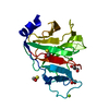
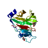
| Structure viewer | Molecule: MolmilJmol/JSmol |
|---|
- Downloads & links
-Download
| PDBx/mmCIF format | 4n7u.cif.gz | 95.8 KB | Display | PDBx/mmCIF format |
|---|---|---|---|---|
| PDB format | pdb4n7u.ent.gz | 72.1 KB | Display | PDB format |
| PDBx/mmJSON format | 4n7u.json.gz | Tree view | PDBx/mmJSON format | |
| Others |  Other downloads Other downloads |
-Validation report
| Arichive directory | https://data.pdbj.org/pub/pdb/validation_reports/n7/4n7uftp://data.pdbj.org/pub/pdb/validation_reports/n7/4n7u | HTTPS FTP |
|---|
-Related structure data
| Related structure data |  4n7iSC S: Starting model for refinement C: citing same article ( |
|---|---|
| Similar structure data |


-Links
 PDBj
PDBj
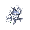
- Assembly
| Deposited unit | 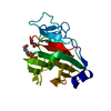
| |||||||||
|---|---|---|---|---|---|---|---|---|---|---|
| 1 |
| |||||||||
| 2 | 
| |||||||||
| Unit cell |
| |||||||||
| Components on special symmetry positions |
|
-Components
| #1: Protein | Mass: 21834.861 Da / Num. of mol.: 1 / Fragment: UNP Residues 328-513 Source method: isolated from a genetically manipulated source Source: (gene. exp.) Homo sapiens (human) / Gene: BTF5, BTN3A1 / Plasmid: pET28a / Production host:  Escherichia coli (E. coli) / Strain (production host): BL21 / References: UniProt: O00481 Escherichia coli (E. coli) / Strain (production host): BL21 / References: UniProt: O00481 |
|---|---|
| #2: Chemical | ChemComp-2JA / [(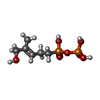  Mass: 260.119 Da / Num. of mol.: 1 / Source method: obtained synthetically / Formula: C6H14O7P2 / Details: Innate Pharma Mass: 260.119 Da / Num. of mol.: 1 / Source method: obtained synthetically / Formula: C6H14O7P2 / Details: Innate Pharma |
| #3: Chemical | ChemComp-GOL / Glycerol  Mass: 92.094 Da / Num. of mol.: 1 / Source method: obtained synthetically / Formula: C3H8O3 Mass: 92.094 Da / Num. of mol.: 1 / Source method: obtained synthetically / Formula: C3H8O3 |
| #4: Water | ChemComp-HOH / Water Mass: 18.015 Da / Num. of mol.: 211 / Source method: isolated from a natural source / Formula: H2O Mass: 18.015 Da / Num. of mol.: 211 / Source method: isolated from a natural source / Formula: H2O |
-Experimental details
-Experiment
| Experiment | Method: X-RAY DIFFRACTION / Number of used crystals: 1 |
|---|
- Sample preparation
| Crystal | Density Matthews: 2.51 Å3/Da / Density % sol: 51.05 % |
|---|---|
| Crystal grow | Temperature: 298 K / Method: vapor diffusion, sitting drop / pH: 7.2 Details: 0.2M MgCl2, 22%PEG3350, pH 7.2, VAPOR DIFFUSION, SITTING DROP, temperature 298K |
-Data collection
| Diffraction | Mean temperature: 100 K | |||||||||
|---|---|---|---|---|---|---|---|---|---|---|
| Diffraction source | Source: SYNCHROTRON / Site: APS  / Beamline: 24-ID-E / Wavelength: 0.97915 Å / Beamline: 24-ID-E / Wavelength: 0.97915 Å | |||||||||
| Detector | Type: ADSC QUANTUM 315 / Detector: CCD / Date: Aug 12, 2013 | |||||||||
| Radiation | Protocol: SINGLE WAVELENGTH / Monochromatic (M) / Laue (L): M / Scattering type: x-ray | |||||||||
| Radiation wavelength | Wavelength: 0.97915 Å / Relative weight: 1 | |||||||||
| Reflection | Resolution: 1.4598→42.31 Å / Num. all: 37813 / Num. obs: 37549 / % possible obs: 96.3 % | |||||||||
| Reflection shell |
|
- Processing
| Software |
| ||||||||||||||||||||||||||||||||||||||||||||||||||||||||||||||||||||||||||||||||||||||||||||||||||
|---|---|---|---|---|---|---|---|---|---|---|---|---|---|---|---|---|---|---|---|---|---|---|---|---|---|---|---|---|---|---|---|---|---|---|---|---|---|---|---|---|---|---|---|---|---|---|---|---|---|---|---|---|---|---|---|---|---|---|---|---|---|---|---|---|---|---|---|---|---|---|---|---|---|---|---|---|---|---|---|---|---|---|---|---|---|---|---|---|---|---|---|---|---|---|---|---|---|---|---|
| Refinement | Method to determine structure: MOLECULAR REPLACEMENT Starting model: PDB ENTRY 4N7I Resolution: 1.4598→37.22 Å / Occupancy max: 1 / Occupancy min: 0.45 / FOM work R set: 0.8823 / SU ML: 0.13 / σ(F): 1.34 / Phase error: 18.53 / Stereochemistry target values: ML
| ||||||||||||||||||||||||||||||||||||||||||||||||||||||||||||||||||||||||||||||||||||||||||||||||||
| Solvent computation | Shrinkage radii: 0.9 Å / VDW probe radii: 1.11 Å / Solvent model: FLAT BULK SOLVENT MODEL | ||||||||||||||||||||||||||||||||||||||||||||||||||||||||||||||||||||||||||||||||||||||||||||||||||
| Displacement parameters | Biso max: 79.84 Å2 / Biso mean: 20.1575 Å2 / Biso min: 6.68 Å2 | ||||||||||||||||||||||||||||||||||||||||||||||||||||||||||||||||||||||||||||||||||||||||||||||||||
| Refinement step | Cycle: LAST / Resolution: 1.4598→37.22 Å
| ||||||||||||||||||||||||||||||||||||||||||||||||||||||||||||||||||||||||||||||||||||||||||||||||||
| Refine LS restraints |
| ||||||||||||||||||||||||||||||||||||||||||||||||||||||||||||||||||||||||||||||||||||||||||||||||||
| LS refinement shell | Refine-ID: X-RAY DIFFRACTION / Total num. of bins used: 13
|
