[English] 日本語
Yorodumi
- PDB-6ip0: Crystal structure of Arabidopsis thaliana JMJ13 catalytic domain ... -

+
Open data


ID or keywords:

Loading...

-
Basic information

Entry
Database: PDB / ID: 6ip0
TitleCrystal structure of Arabidopsis thaliana JMJ13 catalytic domain in complex with AKG
ComponentsTranscription factor jumonji (Jmj) family protein
KeywordsGENE REGULATION / Arabidopsis / histone modification / flowering / epigenetics / H3K27me3
Function / homology
Function and homology information


negative regulation of short-day photoperiodism, flowering / long-day photoperiodism / negative regulation of long-day photoperiodism, flowering / histone H3K27me2/H3K27me3 demethylase activity / flower development / histone H3K4me/H3K4me2/H3K4me3 demethylase activity / Oxidoreductases; Acting on paired donors, with incorporation or reduction of molecular oxygen; With 2-oxoglutarate as one donor, and incorporation of one atom of oxygen into each donor / negative regulation of gene expression, epigenetic / response to temperature stimulus / histone demethylase activity ...negative regulation of short-day photoperiodism, flowering / long-day photoperiodism / negative regulation of long-day photoperiodism, flowering / histone H3K27me2/H3K27me3 demethylase activity / flower development / histone H3K4me/H3K4me2/H3K4me3 demethylase activity / Oxidoreductases; Acting on paired donors, with incorporation or reduction of molecular oxygen; With 2-oxoglutarate as one donor, and incorporation of one atom of oxygen into each donor / negative regulation of gene expression, epigenetic / response to temperature stimulus / histone demethylase activity / histone binding / chromatin remodeling / DNA-binding transcription factor activity / regulation of DNA-templated transcription / metal ion binding / nucleus
Similarity search - Function
Zinc finger, C5HC2-type / C5HC2 zinc finger / JmjN domain / jmjN domain / JmjN domain profile. / Small domain found in the jumonji family of transcription factors / JmjC domain, hydroxylase / A domain family that is part of the cupin metalloenzyme superfamily. / JmjC domain / JmjC domain profile.
Similarity search - Domain/homology
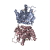
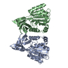
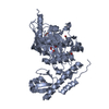
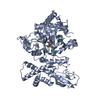
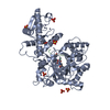
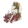
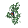
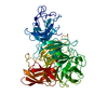
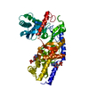
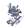
2-OXOGLUTARIC ACID / NICKEL (II) ION / Transcription factor jumonji (Jmj) family protein / zinc finger (C5HC2 type) family protein / Lysine-specific demethylase JMJ13
Similarity search - Component
Biological speciesArabidopsis thaliana (thale cress)
MethodX-RAY DIFFRACTION / SYNCHROTRON / SAD / Resolution: 2.4 Å
AuthorsHu, H. / Du, J.
CitationJournal: Nat Commun / Year: 2019
Title: The Arabidopsis H3K27me3 demethylase JUMONJI 13 is a temperature and photoperiod dependent flowering repressor.
Authors: Zheng, S. / Hu, H. / Ren, H. / Yang, Z. / Qiu, Q. / Qi, W. / Liu, X. / Chen, X. / Cui, X. / Li, S. / Zhou, B. / Sun, D. / Cao, X. / Du, J.
History
DepositionNov 1, 2018Deposition site: PDBJ / Processing site: PDBJ
Revision 1.0Apr 10, 2019Provider: repository / Type: Initial release
Revision 1.1Mar 27, 2024Group: Data collection / Database references / Category: chem_comp_atom / chem_comp_bond / database_2
Item: _database_2.pdbx_DOI / _database_2.pdbx_database_accession

-
Structure visualization

Structure viewerMolecule:
MolmilJmol/JSmol

Downloads & links

-
Assembly

Deposited unit
A: Transcription factor jumonji (Jmj) family protein
hetero molecules


Theoretical massNumber of molelcules
Total (without water)56,91918
Polymers55,3351
Non-polymers1,58417
Water2,630146
1


  • Idetical with deposited unit
  • defined by author
  • Evidence: gel filtration
TypeNameSymmetry operationNumber
identity operation1_555x,y,z1
Buried area2730 Å2
ΔGint-173 kcal/mol
Surface area22110 Å2
Unit cell
Length a, b, c (Å)129.670, 129.670, 231.520
Angle α, β, γ (deg.)90.00, 90.00, 120.00
Int Tables number180
Space group name H-MP6222
Components on special symmetry positions
IDModelComponents
11A-612-

SO4

-
Components

-
Protein , 1 types, 1 molecules A

#1: Protein Transcription factor jumonji (Jmj) family protein / JMJ13 catalytic domain


Mass: 55335.070 Da / Num. of mol.: 1 / Fragment: UNP residues 90-578
Source method: isolated from a genetically manipulated source
Source: (gene. exp.) Arabidopsis thaliana (thale cress) / Plasmid: psumo / Production host: Escherichia coli BL21(DE3) (bacteria) / Strain (production host): BL21(DE3) / Variant (production host): RIL / References: UniProt: A0A1P8BAS6, UniProt: F4KIX0*PLUS

-
Non-polymers , 5 types, 163 molecules

#2: Chemical ChemComp-AKG / 2-OXOGLUTARIC ACID / Α-Ketoglutaric acid


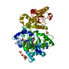
Mass: 146.098 Da / Num. of mol.: 1 / Source method: obtained synthetically / Formula: C5H6O5
#3: Chemical ChemComp-NI / NICKEL (II) ION / Nickel


Mass: 58.693 Da / Num. of mol.: 1 / Source method: obtained synthetically / Formula: Ni
#4: Chemical ChemComp-ZN / ZINC ION


Mass: 65.409 Da / Num. of mol.: 2 / Source method: obtained synthetically / Formula: Zn
#5: Chemical
ChemComp-SO4 / SULFATE ION / Sulfate


Mass: 96.063 Da / Num. of mol.: 13 / Source method: obtained synthetically / Formula: SO4
#6: Water ChemComp-HOH / water / Water


Mass: 18.015 Da / Num. of mol.: 146 / Source method: isolated from a natural source / Formula: H2O

-
Experimental details

-
Experiment

ExperimentMethod: X-RAY DIFFRACTION / Number of used crystals: 1

-
Sample preparation

CrystalDensity Matthews: 5.08 Å3/Da / Density % sol: 75.77 %
Crystal growTemperature: 277 K / Method: vapor diffusion, sitting drop
Details: 0.1M MES, pH 6.5, 8% dioxane, and 1.6M ammonium sulfate

-
Data collection

DiffractionMean temperature: 100 K / Serial crystal experiment: N
Diffraction sourceSource: SYNCHROTRON / Site: SSRF / Beamline: BL19U1 / Wavelength: 1.2824 Å
DetectorType: DECTRIS PILATUS3 S 6M / Detector: PIXEL / Date: Jan 2, 2017
RadiationProtocol: SINGLE WAVELENGTH / Monochromatic (M) / Laue (L): M / Scattering type: x-ray
Radiation wavelengthWavelength: 1.2824 Å / Relative weight: 1
ReflectionResolution: 2.4→50 Å / Num. obs: 45790 / % possible obs: 100 % / Redundancy: 19.3 % / Rmerge(I) obs: 0.078 / Net I/σ(I): 36.15
Reflection shellResolution: 2.4→2.49 Å / Redundancy: 19.4 % / Rmerge(I) obs: 0.651 / Mean I/σ(I) obs: 4 / Num. unique obs: 4476 / CC1/2: 0.945 / Rpim(I) all: 0.151 / Rrim(I) all: 0.668 / Rsym value: 0.651 / % possible all: 100

-
Processing

Software
NameVersionClassification
PHENIX(1.12_2829: ???)refinement
HKL-3000data reduction
HKL-3000data scaling
PHENIXphasing
RefinementMethod to determine structure: SAD / Resolution: 2.4→49.641 Å / SU ML: 0.28 / Cross valid method: FREE R-VALUE / σ(F): 1.36 / Phase error: 21.19
RfactorNum. reflection% reflection
Rfree0.2124 2377 5.19 %
Rwork0.1915 --
obs0.1926 45790 99.96 %
Solvent computationShrinkage radii: 0.9 Å / VDW probe radii: 1.11 Å
Refinement stepCycle: LAST / Resolution: 2.4→49.641 Å
ProteinNucleic acidLigandSolventTotal
Num. atoms3739 0 78 146 3963
Refine LS restraints
Refine-IDTypeDev idealNumber
X-RAY DIFFRACTIONf_bond_d0.0073901
X-RAY DIFFRACTIONf_angle_d0.965294
X-RAY DIFFRACTIONf_dihedral_angle_d22.5011406
X-RAY DIFFRACTIONf_chiral_restr0.065566
X-RAY DIFFRACTIONf_plane_restr0.007665
LS refinement shell
Resolution (Å)Rfactor RfreeNum. reflection RfreeRfactor RworkNum. reflection RworkRefine-ID% reflection obs (%)
2.3974-2.44640.30991390.26692494X-RAY DIFFRACTION100
2.4464-2.49960.28221320.24582504X-RAY DIFFRACTION100
2.4996-2.55770.24721400.21932486X-RAY DIFFRACTION100
2.5577-2.62170.28071370.21872509X-RAY DIFFRACTION100
2.6217-2.69250.24051320.20842509X-RAY DIFFRACTION100
2.6925-2.77180.27581380.21632513X-RAY DIFFRACTION100
2.7718-2.86120.2311270.21822531X-RAY DIFFRACTION100
2.8612-2.96350.21081440.21742516X-RAY DIFFRACTION100
2.9635-3.08210.29411510.22582523X-RAY DIFFRACTION100
3.0821-3.22240.21941460.21292520X-RAY DIFFRACTION100
3.2224-3.39220.27581330.20862558X-RAY DIFFRACTION100
3.3922-3.60470.20711360.1982557X-RAY DIFFRACTION100
3.6047-3.88290.22881440.19222544X-RAY DIFFRACTION100
3.8829-4.27350.1591450.16032578X-RAY DIFFRACTION100
4.2735-4.89140.16531230.14822624X-RAY DIFFRACTION100
4.8914-6.16080.18741640.18922622X-RAY DIFFRACTION100
6.1608-49.65210.19871460.18462825X-RAY DIFFRACTION100
Refinement TLS params.

Method: refined / Refine-ID: X-RAY DIFFRACTION

IDL112)L122)L132)L222)L232)L332)S11 (Å °)S12 (Å °)S13 (Å °)S21 (Å °)S22 (Å °)S23 (Å °)S31 (Å °)S32 (Å °)S33 (Å °)T112)T122)T132)T222)T232)T332)Origin x (Å)Origin y (Å)Origin z (Å)
11.1705-0.8057-0.37012.58010.70771.7292-0.0746-0.15370.02510.10850.0435-0.09550.10010.11580.01960.53250.1987-0.01120.32490.01120.449841.963445.0131142.9468
21.2305-2.1505-0.9224.55471.61790.9530.0542-0.02160.2446-0.40910.0168-0.3591-0.09210.027-0.07170.63670.16680.05250.33440.04270.444753.903435.8479129.9768
34.0807-0.1332.05081.26090.65193.6172-0.1863-0.1473-0.0560.0511-0.04550.1028-0.3311-0.02160.24510.66790.16520.06040.2350.01760.498163.042812.7644130.4459
Refinement TLS group
IDRefine-IDRefine TLS-IDSelection details
1X-RAY DIFFRACTION1chain 'A' and (resid 93 through 317 )
2X-RAY DIFFRACTION2chain 'A' and (resid 318 through 484 )
3X-RAY DIFFRACTION3chain 'A' and (resid 485 through 564 )

+
About Yorodumi

-
News

-
Feb 9, 2022. New format data for meta-information of EMDB entries

New format data for meta-information of EMDB entries

  • Version 3 of the EMDB header file is now the official format.
  • The previous official version 1.9 will be removed from the archive.

Related info.:EMDB header

External links:wwPDB to switch to version 3 of the EMDB data model

-
Aug 12, 2020. Covid-19 info

Covid-19 info

URL: https://pdbj.org/emnavi/covid19.php

New page: Covid-19 featured information page in EM Navigator.

Related info.:Covid-19 info / Mar 5, 2020. Novel coronavirus structure data

+
Mar 5, 2020. Novel coronavirus structure data

Novel coronavirus structure data

Related info.:Yorodumi Speices / Aug 12, 2020. Covid-19 info

External links:COVID-19 featured content - PDBj / Molecule of the Month (242):Coronavirus Proteases

+
Jan 31, 2019. EMDB accession codes are about to change! (news from PDBe EMDB page)

EMDB accession codes are about to change! (news from PDBe EMDB page)

  • The allocation of 4 digits for EMDB accession codes will soon come to an end. Whilst these codes will remain in use, new EMDB accession codes will include an additional digit and will expand incrementally as the available range of codes is exhausted. The current 4-digit format prefixed with “EMD-” (i.e. EMD-XXXX) will advance to a 5-digit format (i.e. EMD-XXXXX), and so on. It is currently estimated that the 4-digit codes will be depleted around Spring 2019, at which point the 5-digit format will come into force.
  • The EM Navigator/Yorodumi systems omit the EMD- prefix.

Related info.:Q: What is EMD? / ID/Accession-code notation in Yorodumi/EM Navigator

External links:EMDB Accession Codes are Changing Soon! / Contact to PDBj

+
Jul 12, 2017. Major update of PDB

Major update of PDB

  • wwPDB released updated PDB data conforming to the new PDBx/mmCIF dictionary.
  • This is a major update changing the version number from 4 to 5, and with Remediation, in which all the entries are updated.
  • In this update, many items about electron microscopy experimental information are reorganized (e.g. em_software).
  • Now, EM Navigator and Yorodumi are based on the updated data.

External links:wwPDB Remediation / Enriched Model Files Conforming to OneDep Data Standards Now Available in the PDB FTP Archive

-
Yorodumi

Thousand views of thousand structures

  • Yorodumi is a browser for structure data from EMDB, PDB, SASBDB, etc.
  • This page is also the successor to EM Navigator detail page, and also detail information page/front-end page for Omokage search.
  • The word "yorodu" (or yorozu) is an old Japanese word meaning "ten thousand". "mi" (miru) is to see.

Related info.:EMDB / PDB / SASBDB / Comparison of 3 databanks / Yorodumi Search / Aug 31, 2016. New EM Navigator & Yorodumi / Yorodumi Papers / Jmol/JSmol / Function and homology information / Changes in new EM Navigator and Yorodumi

Read more