 Movie
Movie Controller

[English] 日本語
 Yorodumi
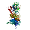
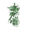
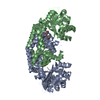
- PDB-6ioi: Crystal structure of Homoserine O-acetyltransferase in complex wi... -
+ Open data

- Basic information
| Entry | Database: PDB / ID: 6ioi | ||||||
|---|---|---|---|---|---|---|---|
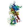
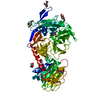
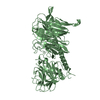
| Title | Crystal structure of Homoserine O-acetyltransferase in complex with CoA from Mycobacterium smegmatis ATCC 19420 | ||||||
 Components Components | Homoserine O-acetyltransferase | ||||||
 Keywords Keywords | TRANSFERASE / BIOSYNTHETIC PROTEIN | ||||||
| Function / homology |  Function and homology informationhomoserine O-acetyltransferase / homoserine O-acetyltransferase activity / methionine biosynthetic process / cytoplasm Function and homology informationhomoserine O-acetyltransferase / homoserine O-acetyltransferase activity / methionine biosynthetic process / cytoplasmSimilarity search - Function | ||||||
| Biological species |  Mycobacterium smegmatis (bacteria) Mycobacterium smegmatis (bacteria) | ||||||
| Method | X-RAY DIFFRACTION / SYNCHROTRON / MOLECULAR REPLACEMENT / Resolution: 1.6 Å | ||||||
 Authors Authors | Sagong, H.-Y. / Kim, K.-J. | ||||||
 Citation Citation | Journal: Biochem.Biophys.Res.Commun. / Year: 2019 Title: Crystal structure and biochemical characterization of O-acetylhomoserine acetyltransferase from Mycobacterium smegmatis ATCC 19420. Authors: Sagong, H.Y. / Hong, J. / Kim, K.J. | ||||||
| History |
|
- Structure visualization
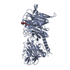
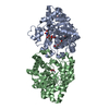
| Structure viewer | Molecule: MolmilJmol/JSmol |
|---|
- Downloads & links
-Download
| PDBx/mmCIF format | 6ioi.cif.gz | 161.5 KB | Display | PDBx/mmCIF format |
|---|---|---|---|---|
| PDB format | pdb6ioi.ent.gz | 126.9 KB | Display | PDB format |
| PDBx/mmJSON format | 6ioi.json.gz | Tree view | PDBx/mmJSON format | |
| Others |  Other downloads Other downloads |
-Validation report
| Arichive directory | https://data.pdbj.org/pub/pdb/validation_reports/io/6ioiftp://data.pdbj.org/pub/pdb/validation_reports/io/6ioi | HTTPS FTP |
|---|
-Related structure data
| Related structure data |  6iogSC 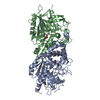 6iohC S: Starting model for refinement C: citing same article ( |
|---|---|
| Similar structure data |

-Links
 PDBj
PDBj


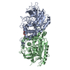
- Assembly
| Deposited unit | 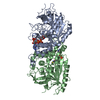
| ||||||||
|---|---|---|---|---|---|---|---|---|---|
| 1 |
| ||||||||
| Unit cell |
|
-Components
| #1: Protein | / HAT / Homoserine transacetylase / HTA Mass: 39475.992 Da / Num. of mol.: 2 Source method: isolated from a genetically manipulated source Source: (gene. exp.) Mycobacterium smegmatis (bacteria) / Gene: metX_1, metXA, ERS451418_01697 / Plasmid: pET30a / Production host: Escherichia coli (E. coli) / Strain (production host): BL21_T1R(DE3)References: UniProt: A0A0D6HE46, UniProt: A0QSZ0*PLUS, homoserine O-acetyltransferase#2: Chemical | Coenzyme A  Mass: 767.534 Da / Num. of mol.: 2 / Source method: obtained synthetically / Formula: C21H36N7O16P3S Mass: 767.534 Da / Num. of mol.: 2 / Source method: obtained synthetically / Formula: C21H36N7O16P3S#3: Water | ChemComp-HOH / | Water Mass: 18.015 Da / Num. of mol.: 577 / Source method: isolated from a natural source / Formula: H2O Mass: 18.015 Da / Num. of mol.: 577 / Source method: isolated from a natural source / Formula: H2O |
|---|
-Experimental details
-Experiment
| Experiment | Method: X-RAY DIFFRACTION / Number of used crystals: 1 |
|---|
- Sample preparation
| Crystal | Density Matthews: 2.56 Å3/Da / Density % sol: 51.99 % |
|---|---|
| Crystal grow | Temperature: 293 K / Method: vapor diffusion, hanging drop / pH: 5.5 Details: 34 % (v/v) polyethylene glycol (PEG) 400, 0.1 M Sodium acetate/acetic acid pH 5.5, 0.2 M calcium acetate |
-Data collection
| Diffraction | Mean temperature: 100 K / Serial crystal experiment: N |
|---|---|
| Diffraction source | Source: SYNCHROTRON / Site: PAL/PLS  / Beamline: 7A (6B, 6C1) / Wavelength: 0.97934 Å / Beamline: 7A (6B, 6C1) / Wavelength: 0.97934 Å |
| Detector | Type: ADSC QUANTUM 270 / Detector: CCD / Date: Oct 24, 2018 |
| Radiation | Monochromator: Double Crystal Monochromator / Protocol: SINGLE WAVELENGTH / Monochromatic (M) / Laue (L): M / Scattering type: x-ray |
| Radiation wavelength | Wavelength: 0.97934 Å / Relative weight: 1 |
| Reflection | Resolution: 1.6→50 Å / Num. obs: 105196 / % possible obs: 99.9 % / Redundancy: 7.2 % / Net I/σ(I): 46 |
| Reflection shell | Resolution: 1.6→1.63 Å / Redundancy: 7.1 % / Mean I/σ(I) obs: 9.56 / Num. unique obs: 5234 / % possible all: 99.9 |
- Processing
| Software |
| ||||||||||||||||||||||||||||||||||||||||||||||||||||||||||||
|---|---|---|---|---|---|---|---|---|---|---|---|---|---|---|---|---|---|---|---|---|---|---|---|---|---|---|---|---|---|---|---|---|---|---|---|---|---|---|---|---|---|---|---|---|---|---|---|---|---|---|---|---|---|---|---|---|---|---|---|---|---|
| Refinement | Method to determine structure: MOLECULAR REPLACEMENT Starting model: 6IOG Resolution: 1.6→30.14 Å / Cor.coef. Fo:Fc: 0.957 / Cor.coef. Fo:Fc free: 0.948 / SU B: 1.39 / SU ML: 0.05 / Cross valid method: THROUGHOUT / σ(F): 0 / ESU R: 0.084 / ESU R Free: 0.084 / Stereochemistry target values: MAXIMUM LIKELIHOOD Details: HYDROGENS HAVE BEEN ADDED IN THE RIDING POSITIONS U VALUES : REFINED INDIVIDUALLY
| ||||||||||||||||||||||||||||||||||||||||||||||||||||||||||||
| Solvent computation | Ion probe radii: 0.8 Å / Shrinkage radii: 0.8 Å / VDW probe radii: 1.2 Å / Solvent model: MASK | ||||||||||||||||||||||||||||||||||||||||||||||||||||||||||||
| Displacement parameters | Biso max: 125.86 Å2 / Biso mean: 16.62 Å2 / Biso min: 8.06 Å2
| ||||||||||||||||||||||||||||||||||||||||||||||||||||||||||||
| Refinement step | Cycle: final / Resolution: 1.6→30.14 Å
| ||||||||||||||||||||||||||||||||||||||||||||||||||||||||||||
| Refine LS restraints |
| ||||||||||||||||||||||||||||||||||||||||||||||||||||||||||||
| LS refinement shell | Resolution: 1.6→1.642 Å / Rfactor Rfree error: 0 / Total num. of bins used: 20
|
