 Movie
Movie Controller


+ Open data

- Basic information
| Entry | Database: PDB / ID: 6in3 | ||||||
|---|---|---|---|---|---|---|---|
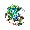
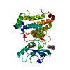
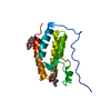
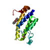
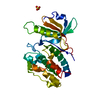
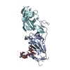
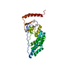
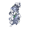
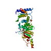
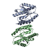
| Title | Crystal structure of DOT1L in complex with 18-Crown-6 | ||||||
 Components Components | Histone-lysine N-methyltransferase, H3 lysine-79 specific Histone methyltransferase Histone methyltransferase | ||||||
 Keywords Keywords | TRANSFERASE / histone H3K79 methyltransferase | ||||||
| Function / homology |  Function and homology information Function and homology informationhistone H3K79 trimethyltransferase activity / [histone H3]-lysine79 N-trimethyltransferase / histone H3K79 methyltransferase activity / regulation of transcription regulatory region DNA binding / histone H3 methyltransferase activity / regulation of receptor signaling pathway via JAK-STAT / histone methyltransferase activity / heterochromatin formation / telomere organization / DNA damage checkpoint signaling ...histone H3K79 trimethyltransferase activity / [histone H3]-lysine79 N-trimethyltransferase / histone H3K79 methyltransferase activity / regulation of transcription regulatory region DNA binding / histone H3 methyltransferase activity / regulation of receptor signaling pathway via JAK-STAT / histone methyltransferase activity / heterochromatin formation / telomere organization / DNA damage checkpoint signaling / PKMTs methylate histone lysines / gene expression / methylation / RNA polymerase II-specific DNA-binding transcription factor binding / nucleic acid binding / transcription coactivator activity / intracellular membrane-bounded organelle / DNA repair / positive regulation of cell population proliferation / positive regulation of transcription by RNA polymerase II / protein-containing complex / DNA binding / nucleoplasm / nucleus / cytoplasmSimilarity search - Function | ||||||
| Biological species |  Homo sapiens (human) Homo sapiens (human) | ||||||
| Method | X-RAY DIFFRACTION / SYNCHROTRON / Resolution: 2.3 Å | ||||||
 Authors Authors | Yokoyama, T. / Kosaka, Y. / Matsumoto, K. / Kitakami, R. / Nabeshima, Y. / Mizuguchi, M. | ||||||
 Citation Citation | Journal: To Be Published Title: Crown Ethers as Transthyretin Amyloidogenesis Inhibitor Authors: Yokoyama, T. / Kosaka, Y. / Matsumoto, K. / Kitakami, R. / Nabeshima, Y. / Mizuguchi, M. | ||||||
| History |
|
- Structure visualization
| Structure viewer | Molecule: MolmilJmol/JSmol |
|---|
- Downloads & links
-Download
| PDBx/mmCIF format | 6in3.cif.gz | 149.3 KB | Display | PDBx/mmCIF format |
|---|---|---|---|---|
| PDB format | pdb6in3.ent.gz | 116.6 KB | Display | PDB format |
| PDBx/mmJSON format | 6in3.json.gz | Tree view | PDBx/mmJSON format | |
| Others |  Other downloads Other downloads |
-Validation report
| Arichive directory | https://data.pdbj.org/pub/pdb/validation_reports/in/6in3ftp://data.pdbj.org/pub/pdb/validation_reports/in/6in3 | HTTPS FTP |
|---|
-Related structure data


-Links
 PDBj
PDBj- Assembly
| Deposited unit | 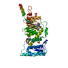
| ||||||||
|---|---|---|---|---|---|---|---|---|---|
| 1 |
| ||||||||
| Unit cell |
|
-Components
| #1: Protein | Histone methyltransferase / DOT1-like protein / Histone H3-K79 methyltransferase / H3-K79-HMTase / Lysine N-methyltransferase 4 Mass: 39050.332 Da / Num. of mol.: 1 Source method: isolated from a genetically manipulated source Source: (gene. exp.) Homo sapiens (human) / Gene: DOT1L, KIAA1814, KMT4 / Production host:  Escherichia coli (E. coli) Escherichia coli (E. coli)References: UniProt: Q8TEK3, histone-lysine N-methyltransferase | ||
|---|---|---|---|
| #2: Chemical | ChemComp-O4B / 18-Crown-6  Mass: 264.315 Da / Num. of mol.: 1 / Source method: obtained synthetically / Formula: C12H24O6 Mass: 264.315 Da / Num. of mol.: 1 / Source method: obtained synthetically / Formula: C12H24O6 | ||
| #3: Chemical | ChemComp-SAH / S-Adenosyl-L-homocysteine  Type: L-peptide linking / Mass: 384.411 Da / Num. of mol.: 1 / Source method: obtained synthetically / Formula: C14H20N6O5S Type: L-peptide linking / Mass: 384.411 Da / Num. of mol.: 1 / Source method: obtained synthetically / Formula: C14H20N6O5S | ||
| #4: Chemical | ChemComp-SO4 / Sulfate  Mass: 96.063 Da / Num. of mol.: 5 / Source method: obtained synthetically / Formula: SO4 Mass: 96.063 Da / Num. of mol.: 5 / Source method: obtained synthetically / Formula: SO4#5: Water | ChemComp-HOH / | Water Mass: 18.015 Da / Num. of mol.: 103 / Source method: isolated from a natural source / Formula: H2O Mass: 18.015 Da / Num. of mol.: 103 / Source method: isolated from a natural source / Formula: H2O |
-Experimental details
-Experiment
| Experiment | Method: X-RAY DIFFRACTION / Number of used crystals: 1 |
|---|
- Sample preparation
| Crystal | Density Matthews: 4.39 Å3/Da / Density % sol: 72.01 % |
|---|---|
| Crystal grow | Temperature: 293 K / Method: vapor diffusion, hanging drop Details: 1.8 M ammonium sulfate, 22 mM acetic acid, 78 mM sodium acetate |
-Data collection
| Diffraction | Mean temperature: 100 K / Serial crystal experiment: N |
|---|---|
| Diffraction source | Source: SYNCHROTRON / Site: Photon Factory  / Beamline: AR-NW12A / Wavelength: 1 Å / Beamline: AR-NW12A / Wavelength: 1 Å |
| Detector | Type: ADSC QUANTUM 210r / Detector: CCD / Date: May 14, 2017 |
| Radiation | Protocol: SINGLE WAVELENGTH / Monochromatic (M) / Laue (L): M / Scattering type: x-ray |
| Radiation wavelength | Wavelength: 1 Å / Relative weight: 1 |
| Reflection | Resolution: 2.3→44.57 Å / Num. obs: 30363 / % possible obs: 99.9 % / Redundancy: 7.5 % / Rpim(I) all: 0.031 / Net I/σ(I): 17.6 |
| Reflection shell | Resolution: 2.3→2.38 Å / Rpim(I) all: 0.354 |
- Processing
| Software |
| ||||||||||||||||||||||||||||||||||||||||||||||||||||||||||||||||||||||||||||||||||||
|---|---|---|---|---|---|---|---|---|---|---|---|---|---|---|---|---|---|---|---|---|---|---|---|---|---|---|---|---|---|---|---|---|---|---|---|---|---|---|---|---|---|---|---|---|---|---|---|---|---|---|---|---|---|---|---|---|---|---|---|---|---|---|---|---|---|---|---|---|---|---|---|---|---|---|---|---|---|---|---|---|---|---|---|---|---|
| Refinement | Resolution: 2.3→42.002 Å / SU ML: 0.27 / Cross valid method: FREE R-VALUE / σ(F): 1.37 / Phase error: 22.47
| ||||||||||||||||||||||||||||||||||||||||||||||||||||||||||||||||||||||||||||||||||||
| Solvent computation | Shrinkage radii: 0.9 Å / VDW probe radii: 1.11 Å | ||||||||||||||||||||||||||||||||||||||||||||||||||||||||||||||||||||||||||||||||||||
| Refinement step | Cycle: LAST / Resolution: 2.3→42.002 Å
| ||||||||||||||||||||||||||||||||||||||||||||||||||||||||||||||||||||||||||||||||||||
| Refine LS restraints |
| ||||||||||||||||||||||||||||||||||||||||||||||||||||||||||||||||||||||||||||||||||||
| LS refinement shell |
| ||||||||||||||||||||||||||||||||||||||||||||||||||||||||||||||||||||||||||||||||||||
| Refinement TLS params. | Method: refined / Origin x: 32.4803 Å / Origin y: 53.8009 Å / Origin z: -5.4368 Å
| ||||||||||||||||||||||||||||||||||||||||||||||||||||||||||||||||||||||||||||||||||||
| Refinement TLS group | Selection details: resseq 1:350 and not resname HOH |
