 Movie
Movie Controller

[English] 日本語
 Yorodumi
- PDB-6i3y: Crystal structure of the human mitochondrial PRELID1K58V-TRIAP1 c... -
+ Open data

- Basic information
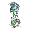
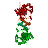
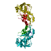
| Entry | Database: PDB / ID: 6i3y | ||||||
|---|---|---|---|---|---|---|---|
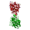
| Title | Crystal structure of the human mitochondrial PRELID1K58V-TRIAP1 complex with PS | ||||||
 Components Components |
| ||||||
 Keywords Keywords | LIPID TRANSPORT / Mitochondrial lipid transport /  Complex / Phosphatidylserine bound / PA transport / Apoptosis Complex / Phosphatidylserine bound / PA transport / Apoptosis | ||||||
| Function / homology |  Function and homology information Function and homology informationregulation of membrane lipid distribution / phosphatidic acid transfer activity / positive regulation of phospholipid transport / positive regulation of endopeptidase activity / positive regulation of cellular respiration / intermembrane lipid transfer / phospholipid transport / positive regulation of T cell apoptotic process / phospholipid translocation / regulation of T cell differentiation ...regulation of membrane lipid distribution / phosphatidic acid transfer activity / positive regulation of phospholipid transport / positive regulation of endopeptidase activity / positive regulation of cellular respiration / intermembrane lipid transfer / phospholipid transport / positive regulation of T cell apoptotic process / phospholipid translocation / regulation of T cell differentiation / negative regulation of intrinsic apoptotic signaling pathway in response to DNA damage by p53 class mediator / negative regulation of release of cytochrome c from mitochondria / negative regulation of mitochondrial membrane potential / TP53 Regulates Transcription of Genes Involved in Cytochrome C Release / DNA damage response, signal transduction by p53 class mediator / Mitochondrial protein degradation / DNA damage response, signal transduction by p53 class mediator resulting in cell cycle arrest / regulation of mitochondrial membrane potential / negative regulation of cysteine-type endopeptidase activity involved in apoptotic process / mitochondrial intermembrane space / cellular response to UV / p53 binding / apoptotic process / negative regulation of apoptotic process / positive regulation of transcription by RNA polymerase II / protein-containing complex / mitochondrion / nucleoplasm / nucleus / cytosolSimilarity search - Function | ||||||
| Biological species |  Homo sapiens (human) Homo sapiens (human) | ||||||
| Method | X-RAY DIFFRACTION / SYNCHROTRON / MOLECULAR REPLACEMENT / Resolution: 2.98 Å | ||||||
 Authors Authors | Miliara, X. / Berry, J.-L. / Morgan, R.M.L. / Matthews, S.J. | ||||||
| Funding support |  United Kingdom, 1items United Kingdom, 1items
| ||||||
 Citation Citation | Journal: Nat Commun / Year: 2019 Title: Structural determinants of lipid specificity within Ups/PRELI lipid transfer proteins. Authors: Miliara, X. / Tatsuta, T. / Berry, J.L. / Rouse, S.L. / Solak, K. / Chorev, D.S. / Wu, D. / Robinson, C.V. / Matthews, S. / Langer, T. | ||||||
| History |
|
- Structure visualization
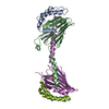
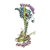
| Structure viewer | Molecule: MolmilJmol/JSmol |
|---|
- Downloads & links
-Download
| PDBx/mmCIF format | 6i3y.cif.gz | 118.4 KB | Display | PDBx/mmCIF format |
|---|---|---|---|---|
| PDB format | pdb6i3y.ent.gz | 87.3 KB | Display | PDB format |
| PDBx/mmJSON format | 6i3y.json.gz | Tree view | PDBx/mmJSON format | |
| Others |  Other downloads Other downloads |
-Validation report
| Arichive directory | https://data.pdbj.org/pub/pdb/validation_reports/i3/6i3yftp://data.pdbj.org/pub/pdb/validation_reports/i3/6i3y | HTTPS FTP |
|---|
-Related structure data
| Related structure data |  6i3vSC  6i4yC S: Starting model for refinement C: citing same article ( |
|---|---|
| Similar structure data |


-Links
 PDBj
PDBj


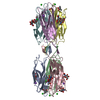
- Assembly
| Deposited unit | 
| ||||||||
|---|---|---|---|---|---|---|---|---|---|
| 1 | 
| ||||||||
| Unit cell |
|
-Components
| #1: Protein | Mass: 21551.279 Da / Num. of mol.: 2 / Mutation: K58V Source method: isolated from a genetically manipulated source Source: (gene. exp.) Homo sapiens (human) / Gene: PRELID1, PRELI, CGI-106, SBBI12 / Plasmid: pET_Duet / Production host:  Escherichia coli (E. coli) / Variant (production host): SHuffle / References: UniProt: Q9Y255 Escherichia coli (E. coli) / Variant (production host): SHuffle / References: UniProt: Q9Y255#2: Protein | Mass: 10518.743 Da / Num. of mol.: 2 Source method: isolated from a genetically manipulated source Source: (gene. exp.) Homo sapiens (human) / Gene: TRIAP1, 15E1.1, HSPC132 / Plasmid: pRSF2 / Production host: Escherichia coli (E. coli) / Variant (production host): SHuffle / References: UniProt: O43715#3: Sugar | ChemComp-LMT / |   Type: D-saccharide / Mass: 510.615 Da / Num. of mol.: 1 / Source method: obtained synthetically / Formula: C24H46O11 / Comment: detergent*YM Type: D-saccharide / Mass: 510.615 Da / Num. of mol.: 1 / Source method: obtained synthetically / Formula: C24H46O11 / Comment: detergent*YM#4: Chemical | Phosphatidylserine  Mass: 792.075 Da / Num. of mol.: 2 / Source method: obtained synthetically / Formula: C42H82NO10P / Feature type: SUBJECT OF INVESTIGATION Mass: 792.075 Da / Num. of mol.: 2 / Source method: obtained synthetically / Formula: C42H82NO10P / Feature type: SUBJECT OF INVESTIGATION#5: Water | ChemComp-HOH / | Water Mass: 18.015 Da / Num. of mol.: 21 / Source method: isolated from a natural source / Formula: H2O Mass: 18.015 Da / Num. of mol.: 21 / Source method: isolated from a natural source / Formula: H2O |
|---|
-Experimental details
-Experiment
| Experiment | Method: X-RAY DIFFRACTION / Number of used crystals: 1 |
|---|
- Sample preparation
| Crystal | Density Matthews: 3.32 Å3/Da / Density % sol: 62.93 % / Description: large hexagonal rods |
|---|---|
| Crystal grow | Temperature: 293.15 K / Method: vapor diffusion, sitting drop / pH: 6.5 / Details: 100 mM sodium cacodylate pH 6.5 40% PEG 300 |
-Data collection
| Diffraction | Mean temperature: 100 K / Serial crystal experiment: N |
|---|---|
| Diffraction source | Source: SYNCHROTRON / Site: Diamond / Beamline: I04 / Wavelength: 0.9795 Å |
| Detector | Type: DECTRIS PILATUS 6M-F / Detector: PIXEL / Date: Jan 31, 2018 |
| Radiation | Protocol: SINGLE WAVELENGTH / Monochromatic (M) / Laue (L): M / Scattering type: x-ray |
| Radiation wavelength | Wavelength: 0.9795 Å / Relative weight: 1 |
| Reflection | Resolution: 2.98→63 Å / Num. obs: 17749 / % possible obs: 100 % / Redundancy: 37.1 % / Net I/σ(I): 9.1 |
| Reflection shell | Resolution: 2.98→3.03 Å / Num. unique obs: 894 / % possible all: 100 |
- Processing
| Software |
| |||||||||||||||||||||||||||||||||||||||||||||||||||||||||||||||||||||||||||||||||||||||||||||||||||||||||||||||||||||||||||||||||||||||||||||||||||||||||||||||||||||||||||||||||||||||||
|---|---|---|---|---|---|---|---|---|---|---|---|---|---|---|---|---|---|---|---|---|---|---|---|---|---|---|---|---|---|---|---|---|---|---|---|---|---|---|---|---|---|---|---|---|---|---|---|---|---|---|---|---|---|---|---|---|---|---|---|---|---|---|---|---|---|---|---|---|---|---|---|---|---|---|---|---|---|---|---|---|---|---|---|---|---|---|---|---|---|---|---|---|---|---|---|---|---|---|---|---|---|---|---|---|---|---|---|---|---|---|---|---|---|---|---|---|---|---|---|---|---|---|---|---|---|---|---|---|---|---|---|---|---|---|---|---|---|---|---|---|---|---|---|---|---|---|---|---|---|---|---|---|---|---|---|---|---|---|---|---|---|---|---|---|---|---|---|---|---|---|---|---|---|---|---|---|---|---|---|---|---|---|---|---|---|---|
| Refinement | Method to determine structure: MOLECULAR REPLACEMENT Starting model: 6I3V Resolution: 2.98→63 Å / Cor.coef. Fo:Fc: 0.942 / Cor.coef. Fo:Fc free: 0.927 / SU B: 28.173 / SU ML: 0.471 / Cross valid method: THROUGHOUT / ESU R: 1.311 / ESU R Free: 0.436 / Details: HYDROGENS HAVE BEEN ADDED IN THE RIDING POSITIONS
| |||||||||||||||||||||||||||||||||||||||||||||||||||||||||||||||||||||||||||||||||||||||||||||||||||||||||||||||||||||||||||||||||||||||||||||||||||||||||||||||||||||||||||||||||||||||||
| Solvent computation | Ion probe radii: 0.8 Å / Shrinkage radii: 0.8 Å / VDW probe radii: 1.2 Å | |||||||||||||||||||||||||||||||||||||||||||||||||||||||||||||||||||||||||||||||||||||||||||||||||||||||||||||||||||||||||||||||||||||||||||||||||||||||||||||||||||||||||||||||||||||||||
| Displacement parameters | Biso mean: 105.079 Å2
| |||||||||||||||||||||||||||||||||||||||||||||||||||||||||||||||||||||||||||||||||||||||||||||||||||||||||||||||||||||||||||||||||||||||||||||||||||||||||||||||||||||||||||||||||||||||||
| Refinement step | Cycle: 1 / Resolution: 2.98→63 Å
| |||||||||||||||||||||||||||||||||||||||||||||||||||||||||||||||||||||||||||||||||||||||||||||||||||||||||||||||||||||||||||||||||||||||||||||||||||||||||||||||||||||||||||||||||||||||||
| Refine LS restraints |
| |||||||||||||||||||||||||||||||||||||||||||||||||||||||||||||||||||||||||||||||||||||||||||||||||||||||||||||||||||||||||||||||||||||||||||||||||||||||||||||||||||||||||||||||||||||||||
| LS refinement shell | Resolution: 2.98→3.057 Å / Total num. of bins used: 20
|
