- PDB-4ytx: Crystal structure of Ups1-Mdm35 complex with PA -
+
Open data
ID or keywords:
Loading...
-
Basic information
Entry
Database: PDB / ID: 4ytx
Title
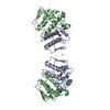
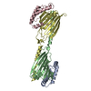
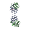
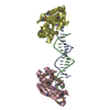
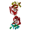
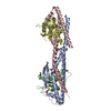
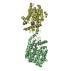
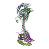
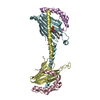
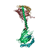
Crystal structure of Ups1-Mdm35 complex with PA
Components
Mitochondrial distribution and morphology protein 35
Protein UPS1, mitochondrial
Keywords
LIPID TRANSPORT / Phospholipid transfer / Mitochondria / Phosphatidic acid
Function / homology
Function and homology information
TP53 Regulates Transcription of Genes Involved in Cytochrome C Release / phosphatidic acid transfer activity / cardiolipin metabolic process / positive regulation of phosphatidylcholine biosynthetic process / mitochondrial respiratory chain complex assembly / phospholipid transport / phospholipid translocation / mitochondrion organization / mitochondrial intermembrane space / mitochondrial inner membrane ...TP53 Regulates Transcription of Genes Involved in Cytochrome C Release / phosphatidic acid transfer activity / cardiolipin metabolic process / positive regulation of phosphatidylcholine biosynthetic process / mitochondrial respiratory chain complex assembly / phospholipid transport / phospholipid translocation / mitochondrion organization / mitochondrial intermembrane space / mitochondrial inner membrane / lipid binding / mitochondrion / nucleus / cytosol / cytoplasm Similarity search - Function
PRELI/MSF1 domain / Slowmo/Ups family / PRELI-like family / PRELI/MSF1 domain profile. / Mitochondrial distribution/morphology family 35/apoptosis / Uncharacterised protein family (UPF0203) / Coiled coil-helix-coiled coil-helix (CHCH) domain profile. Similarity search - Domain/homology
1,2-DILAUROYL-SN-GLYCERO-3-PHOSPHATE / Mitochondrial distribution and morphology protein 35 / Protein UPS1, mitochondrial Similarity search - Component
A: Mitochondrial distribution and morphology protein 35 B: Protein UPS1, mitochondrial C: Mitochondrial distribution and morphology protein 35 D: Protein UPS1, mitochondrial E: Mitochondrial distribution and morphology protein 35 F: Protein UPS1, mitochondrial G: Mitochondrial distribution and morphology protein 35 H: Protein UPS1, mitochondrial I: Mitochondrial distribution and morphology protein 35 J: Protein UPS1, mitochondrial K: Mitochondrial distribution and morphology protein 35 L: Protein UPS1, mitochondrial M: Mitochondrial distribution and morphology protein 35 N: Protein UPS1, mitochondrial O: Mitochondrial distribution and morphology protein 35 P: Protein UPS1, mitochondrial hetero molecules
A: Mitochondrial distribution and morphology protein 35 B: Protein UPS1, mitochondrial M: Mitochondrial distribution and morphology protein 35 N: Protein UPS1, mitochondrial hetero molecules
C: Mitochondrial distribution and morphology protein 35 D: Protein UPS1, mitochondrial E: Mitochondrial distribution and morphology protein 35 F: Protein UPS1, mitochondrial hetero molecules
G: Mitochondrial distribution and morphology protein 35 H: Protein UPS1, mitochondrial O: Mitochondrial distribution and morphology protein 35 P: Protein UPS1, mitochondrial
I: Mitochondrial distribution and morphology protein 35 J: Protein UPS1, mitochondrial K: Mitochondrial distribution and morphology protein 35 L: Protein UPS1, mitochondrial
Chain B and N form a domain-swapped dimer because of the crystallization artifact. The chain B(1-134) and N(135-169) comprise one molecule. The chain N(1-134) and B(135-169) comprise one molecule. The biological assembly is two dimers #1 chain A and B(1-134)/N(135-169), #2 chain M and N(1-134)/B(135-169). The other chains (C,E,D,F), chains (I,J,K,L), chains (G,O,H,P) have the same situation with #1 and #2.
In the structure databanks used in Yorodumi, some data are registered as the other names, "COVID-19 virus" and "2019-nCoV". Here are the details of the virus and the list of structure data.
Jan 31, 2019. EMDB accession codes are about to change! (news from PDBe EMDB page)
EMDB accession codes are about to change! (news from PDBe EMDB page)
The allocation of 4 digits for EMDB accession codes will soon come to an end. Whilst these codes will remain in use, new EMDB accession codes will include an additional digit and will expand incrementally as the available range of codes is exhausted. The current 4-digit format prefixed with “EMD-” (i.e. EMD-XXXX) will advance to a 5-digit format (i.e. EMD-XXXXX), and so on. It is currently estimated that the 4-digit codes will be depleted around Spring 2019, at which point the 5-digit format will come into force.
The EM Navigator/Yorodumi systems omit the EMD- prefix.
Related info.:Q: What is EMD? / ID/Accession-code notation in Yorodumi/EM Navigator
Yorodumi is a browser for structure data from EMDB, PDB, SASBDB, etc.
This page is also the successor to EM Navigator detail page, and also detail information page/front-end page for Omokage search.
The word "yorodu" (or yorozu) is an old Japanese word meaning "ten thousand". "mi" (miru) is to see.
Related info.:EMDB / PDB / SASBDB / Comparison of 3 databanks / Yorodumi Search / Aug 31, 2016. New EM Navigator & Yorodumi / Yorodumi Papers / Jmol/JSmol / Function and homology information / Changes in new EM Navigator and Yorodumi
 Movie
Movie Controller


 Open data
 Basic information
 Components
 Keywords
 Mitochondria /
  Function and homology information
 Authors
 Citation
 Structure visualization
 Downloads & links
 Other downloads
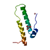


 PDBj
PDBj

 Assembly


 Mass: 535.671 Da / Num. of mol.: 2 / Source method: obtained synthetically / Formula: C27H52O8P
 Sample preparation
 / Beamline: AR-NW12A / Wavelength: 1 Å
 Processing