 Movie
Movie Controller

[English] 日本語
 Yorodumi
- PDB-6hm4: Crystal structure of Rad4 BRCT1,2 in complex with a Mdb1 phosphop... -
+ Open data

- Basic information
| Entry | Database: PDB / ID: 6hm4 | ||||||
|---|---|---|---|---|---|---|---|
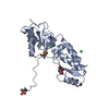
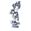
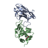
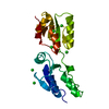
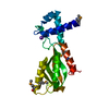
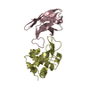
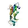
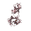
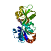
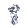
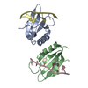
| Title | Crystal structure of Rad4 BRCT1,2 in complex with a Mdb1 phosphopeptide | ||||||
 Components Components |
| ||||||
 Keywords Keywords |  CELL CYCLE / BRCT domain phosphopeptide recognition CELL CYCLE / BRCT domain phosphopeptide recognition | ||||||
| Function / homology |  Function and homology information Function and homology informationmitotic DNA damage checkpoint signaling / mitotic spindle pole body / mitotic spindle midzone / DNA replication preinitiation complex / mitotic DNA replication checkpoint signaling / nuclear replication fork / mitotic G2 DNA damage checkpoint signaling / DNA replication initiation / signaling adaptor activity / mitotic spindle ...mitotic DNA damage checkpoint signaling / mitotic spindle pole body / mitotic spindle midzone / DNA replication preinitiation complex / mitotic DNA replication checkpoint signaling / nuclear replication fork / mitotic G2 DNA damage checkpoint signaling / DNA replication initiation / signaling adaptor activity / mitotic spindle / site of double-strand break / chromatin / nucleus / cytosol / cytoplasmSimilarity search - Function | ||||||
| Biological species |  Schizosaccharomyces pombe (fission yeast) Schizosaccharomyces pombe (fission yeast) | ||||||
| Method | X-RAY DIFFRACTION / SYNCHROTRON / Resolution: 1.77018592355 Å | ||||||
 Authors Authors | Day, M. / Rappas, M. / Oliver, A.W. / Pearl, L.H. | ||||||
| Funding support |  United Kingdom, 1items United Kingdom, 1items
| ||||||
 Citation Citation | Journal: Elife / Year: 2018 Title: BRCT domains of the DNA damage checkpoint proteins TOPBP1/Rad4 display distinct specificities for phosphopeptide ligands. Authors: Day, M. / Rappas, M. / Ptasinska, K. / Boos, D. / Oliver, A.W. / Pearl, L.H. | ||||||
| History |
|
- Structure visualization
| Structure viewer | Molecule: MolmilJmol/JSmol |
|---|
- Downloads & links
-Download
| PDBx/mmCIF format | 6hm4.cif.gz | 68.2 KB | Display | PDBx/mmCIF format |
|---|---|---|---|---|
| PDB format | pdb6hm4.ent.gz | 42.7 KB | Display | PDB format |
| PDBx/mmJSON format | 6hm4.json.gz | Tree view | PDBx/mmJSON format | |
| Others |  Other downloads Other downloads |
-Validation report
| Arichive directory | https://data.pdbj.org/pub/pdb/validation_reports/hm/6hm4ftp://data.pdbj.org/pub/pdb/validation_reports/hm/6hm4 | HTTPS FTP |
|---|
-Related structure data

-Links
 PDBj
PDBj

- Assembly
| Deposited unit | 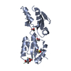
| ||||||||||||
|---|---|---|---|---|---|---|---|---|---|---|---|---|---|
| 1 |
| ||||||||||||
| Unit cell |
|
-Components
| #1: Protein | Mass: 21286.475 Da / Num. of mol.: 1 Source method: isolated from a genetically manipulated source Source: (gene. exp.) Schizosaccharomyces pombe (strain 972 / ATCC 24843) (yeast)Strain: 972 / ATCC 24843 / Gene: rad4, cut5, SPAC23C4.18c / Production host:  Escherichia coli BL21(DE3) (bacteria) / References: UniProt: P32372 Escherichia coli BL21(DE3) (bacteria) / References: UniProt: P32372 |
|---|---|
| #2: Protein/peptide | Mass: 1704.857 Da / Num. of mol.: 1 / Source method: obtained synthetically / Source: (synth.) Schizosaccharomyces pombe (fission yeast) / References: UniProt: O14079 |
| #3: Chemical | ChemComp-ACT / Acetate  Mass: 59.044 Da / Num. of mol.: 1 / Source method: obtained synthetically / Formula: C2H3O2 Mass: 59.044 Da / Num. of mol.: 1 / Source method: obtained synthetically / Formula: C2H3O2 |
| #4: Chemical | ChemComp-EDO / Ethylene glycol  Mass: 62.068 Da / Num. of mol.: 1 / Source method: obtained synthetically / Formula: C2H6O2 Mass: 62.068 Da / Num. of mol.: 1 / Source method: obtained synthetically / Formula: C2H6O2 |
| #5: Water | ChemComp-HOH / Water Mass: 18.015 Da / Num. of mol.: 288 / Source method: isolated from a natural source / Formula: H2O Mass: 18.015 Da / Num. of mol.: 288 / Source method: isolated from a natural source / Formula: H2O |
-Experimental details
-Experiment
| Experiment | Method: X-RAY DIFFRACTION / Number of used crystals: 1 |
|---|
- Sample preparation
| Crystal | Density Matthews: 2.43 Å3/Da / Density % sol: 49.35 % |
|---|---|
| Crystal grow | Temperature: 287.15 K / Method: vapor diffusion, sitting drop Details: 200 mM Sodium acetate trihydrate, 100 mM Tris 8.5 and 30 % w/v PEG 4000 |
-Data collection
| Diffraction | Mean temperature: 100 K |
|---|---|
| Diffraction source | Source: SYNCHROTRON / Site: Diamond / Beamline: I02 / Wavelength: 0.9795 Å |
| Detector | Type: DECTRIS PILATUS 6M-F / Detector: PIXEL / Date: Oct 11, 2014 |
| Radiation | Protocol: SINGLE WAVELENGTH / Monochromatic (M) / Laue (L): M / Scattering type: x-ray |
| Radiation wavelength | Wavelength: 0.9795 Å / Relative weight: 1 |
| Reflection | Resolution: 1.77→31.94 Å / Num. obs: 19339 / % possible obs: 93.07 % / Redundancy: 2.6 % / Biso Wilson estimate: 20.48 Å2 / Net I/σ(I): 16.52 |
| Reflection shell | Resolution: 1.77→1.833 Å |
- Processing
| Software |
| ||||||||||||||||||||||||||||||||||||||||||||||||||||||||
|---|---|---|---|---|---|---|---|---|---|---|---|---|---|---|---|---|---|---|---|---|---|---|---|---|---|---|---|---|---|---|---|---|---|---|---|---|---|---|---|---|---|---|---|---|---|---|---|---|---|---|---|---|---|---|---|---|---|
| Refinement | Resolution: 1.77018592355→31.9379091525 Å / SU ML: 0.232410282223 / Cross valid method: FREE R-VALUE / σ(F): 1.36458498044 / Phase error: 22.5724322855
| ||||||||||||||||||||||||||||||||||||||||||||||||||||||||
| Solvent computation | Shrinkage radii: 0.9 Å / VDW probe radii: 1.11 Å | ||||||||||||||||||||||||||||||||||||||||||||||||||||||||
| Displacement parameters | Biso mean: 22.6342605533 Å2 | ||||||||||||||||||||||||||||||||||||||||||||||||||||||||
| Refinement step | Cycle: LAST / Resolution: 1.77018592355→31.9379091525 Å
| ||||||||||||||||||||||||||||||||||||||||||||||||||||||||
| Refine LS restraints |
| ||||||||||||||||||||||||||||||||||||||||||||||||||||||||
| LS refinement shell |
|
