 Movie
Movie Controller


+ Open data

- Basic information
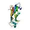
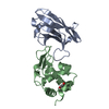
| Entry | Database: PDB / ID: 6ewg | ||||||
|---|---|---|---|---|---|---|---|
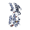
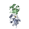
| Title | Oreochromis niloticus CEP120 second C2 domain (C2B) | ||||||
 Components Components | Centrosomal protein 120 Centrosome Centrosome | ||||||
 Keywords Keywords | CYTOSOLIC PROTEIN / Centriole Centrosome Basal body Cilia | ||||||
| Function / homology | Domain of unknown function DUF3668 / Centrosomal protein of 120kDa-like / Cep120 protein / C2 domain / C2 domain / C2 domain profile. / C2 domain superfamily / Centrosomal protein 120 Function and homology information Function and homology information | ||||||
| Biological species |  Oreochromis niloticus (Nile tilapia) Oreochromis niloticus (Nile tilapia) | ||||||
| Method | X-RAY DIFFRACTION / SYNCHROTRON / MOLECULAR REPLACEMENT / Resolution: 1.6 Å | ||||||
 Authors Authors | van Breugel, M. | ||||||
| Funding support |  United Kingdom, 1items United Kingdom, 1items
| ||||||
 Citation Citation | Journal: Cell Rep / Year: 2018 Title: Disease-Associated Mutations in CEP120 Destabilize the Protein and Impair Ciliogenesis. Authors: Joseph, N. / Al-Jassar, C. / Johnson, C.M. / Andreeva, A. / Barnabas, D.D. / Freund, S.M.V. / Gergely, F. / van Breugel, M. | ||||||
| History |
|
- Structure visualization
| Structure viewer | Molecule: MolmilJmol/JSmol |
|---|
- Downloads & links
-Download
| PDBx/mmCIF format | 6ewg.cif.gz | 93.2 KB | Display | PDBx/mmCIF format |
|---|---|---|---|---|
| PDB format | pdb6ewg.ent.gz | 70.2 KB | Display | PDB format |
| PDBx/mmJSON format | 6ewg.json.gz | Tree view | PDBx/mmJSON format | |
| Others |  Other downloads Other downloads |
-Validation report
| Arichive directory | https://data.pdbj.org/pub/pdb/validation_reports/ew/6ewgftp://data.pdbj.org/pub/pdb/validation_reports/ew/6ewg | HTTPS FTP |
|---|
-Related structure data
| Related structure data |  6ewhSC  6ewiC  6ewlC  6ewpC S: Starting model for refinement C: citing same article ( |
|---|---|
| Similar structure data |


-Links
 PDBj
PDBj


- Assembly
| Deposited unit | 
| ||||||||
|---|---|---|---|---|---|---|---|---|---|
| 1 |
| ||||||||
| Unit cell |
|
-Components
| #1: Protein | Centrosome Mass: 20994.924 Da / Num. of mol.: 1 Source method: isolated from a genetically manipulated source Source: (gene. exp.) Oreochromis niloticus (Nile tilapia) / Gene: cep120 / Production host:  Escherichia coli (E. coli) / Strain (production host): Rosetta / References: UniProt: I3K8D3 Escherichia coli (E. coli) / Strain (production host): Rosetta / References: UniProt: I3K8D3 |
|---|---|
| #2: Water | ChemComp-HOH / Water Mass: 18.015 Da / Num. of mol.: 142 / Source method: isolated from a natural source / Formula: H2O Mass: 18.015 Da / Num. of mol.: 142 / Source method: isolated from a natural source / Formula: H2O |
-Experimental details
-Experiment
| Experiment | Method: X-RAY DIFFRACTION / Number of used crystals: 1 |
|---|
- Sample preparation
| Crystal | Density Matthews: 2.61 Å3/Da / Density % sol: 52.8 % |
|---|---|
| Crystal grow | Temperature: 292 K / Method: vapor diffusion, sitting drop / pH: 6.5 / Details: 0.1 Na-MES pH ~6.5 30% PEG 400 / PH range: pH gradient around pH 6.5 |
-Data collection
| Diffraction | Mean temperature: 100 K |
|---|---|
| Diffraction source | Source: SYNCHROTRON / Site: Diamond / Beamline: I04 / Wavelength: 0.9795 Å |
| Detector | Type: DECTRIS PILATUS 6M-F / Detector: PIXEL / Date: Jan 16, 2017 |
| Radiation | Protocol: SINGLE WAVELENGTH / Monochromatic (M) / Laue (L): M / Scattering type: x-ray |
| Radiation wavelength | Wavelength: 0.9795 Å / Relative weight: 1 |
| Reflection | Resolution: 1.6→89.12 Å / Num. obs: 29804 / % possible obs: 100 % / Redundancy: 12.7 % / Biso Wilson estimate: 28.5 Å2 / CC1/2: 0.999 / Rmerge(I) obs: 0.062 / Rpim(I) all: 0.018 / Rrim(I) all: 0.065 / Net I/σ(I): 15.5 |
| Reflection shell | Resolution: 1.6→1.63 Å / Redundancy: 13.2 % / Mean I/σ(I) obs: 1.6 / Num. measured obs: 29602 / Num. unique obs: 1461 / CC1/2: 0.812 / Rpim(I) all: 0.536 / % possible all: 100 |
- Processing
| Software |
| |||||||||||||||||||||||||||||||||||||||||||||||||||||||||||||||||||||||||||||||||||||||||||||||||||||||||||||||||||||||||||||||||||||||||||||||||||
|---|---|---|---|---|---|---|---|---|---|---|---|---|---|---|---|---|---|---|---|---|---|---|---|---|---|---|---|---|---|---|---|---|---|---|---|---|---|---|---|---|---|---|---|---|---|---|---|---|---|---|---|---|---|---|---|---|---|---|---|---|---|---|---|---|---|---|---|---|---|---|---|---|---|---|---|---|---|---|---|---|---|---|---|---|---|---|---|---|---|---|---|---|---|---|---|---|---|---|---|---|---|---|---|---|---|---|---|---|---|---|---|---|---|---|---|---|---|---|---|---|---|---|---|---|---|---|---|---|---|---|---|---|---|---|---|---|---|---|---|---|---|---|---|---|---|---|---|---|
| Refinement | Method to determine structure: MOLECULAR REPLACEMENT Starting model: 6EWH Resolution: 1.6→37.186 Å / SU ML: 0.19 / Cross valid method: FREE R-VALUE / σ(F): 0 / Phase error: 25.77
| |||||||||||||||||||||||||||||||||||||||||||||||||||||||||||||||||||||||||||||||||||||||||||||||||||||||||||||||||||||||||||||||||||||||||||||||||||
| Solvent computation | Shrinkage radii: 0.9 Å / VDW probe radii: 1.11 Å | |||||||||||||||||||||||||||||||||||||||||||||||||||||||||||||||||||||||||||||||||||||||||||||||||||||||||||||||||||||||||||||||||||||||||||||||||||
| Refinement step | Cycle: LAST / Resolution: 1.6→37.186 Å
| |||||||||||||||||||||||||||||||||||||||||||||||||||||||||||||||||||||||||||||||||||||||||||||||||||||||||||||||||||||||||||||||||||||||||||||||||||
| Refine LS restraints |
| |||||||||||||||||||||||||||||||||||||||||||||||||||||||||||||||||||||||||||||||||||||||||||||||||||||||||||||||||||||||||||||||||||||||||||||||||||
| LS refinement shell |
| |||||||||||||||||||||||||||||||||||||||||||||||||||||||||||||||||||||||||||||||||||||||||||||||||||||||||||||||||||||||||||||||||||||||||||||||||||
| Refinement TLS params. | Method: refined / Origin x: -4.8255 Å / Origin y: 7.848 Å / Origin z: -14.9286 Å
| |||||||||||||||||||||||||||||||||||||||||||||||||||||||||||||||||||||||||||||||||||||||||||||||||||||||||||||||||||||||||||||||||||||||||||||||||||
| Refinement TLS group | Selection details: all |
