 Movie
Movie Controller

[English] 日本語
 Yorodumi
- PDB-6h48: A polyamorous repressor: deciphering the evolutionary strategy us... -
+ Open data

- Basic information
| Entry | Database: PDB / ID: 6h48 | |||||||||||||||||||||
|---|---|---|---|---|---|---|---|---|---|---|---|---|---|---|---|---|---|---|---|---|---|---|
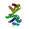
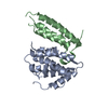
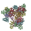
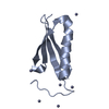
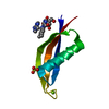
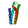
| Title | A polyamorous repressor: deciphering the evolutionary strategy used by the phage-inducible chromosomal islands to spread in nature. | |||||||||||||||||||||
 Components Components | Orf20 | |||||||||||||||||||||
 Keywords Keywords |  STRUCTURAL PROTEIN / SaPI / repressor STRUCTURAL PROTEIN / SaPI / repressor | |||||||||||||||||||||
| Function / homology | Helix-turn-helix / Helix-turn-helix XRE-family like proteins / Cro/C1-type HTH domain profile. / Cro/C1-type helix-turn-helix domain / Lambda repressor-like, DNA-binding domain superfamily / DNA binding / XRE family transcriptional regulator / Orf20 Function and homology information Function and homology information | |||||||||||||||||||||
| Biological species |   Staphylococcus aureus (bacteria) Staphylococcus aureus (bacteria) | |||||||||||||||||||||
| Method | X-RAY DIFFRACTION / SYNCHROTRON / SAD / Resolution: 2.2 Å | |||||||||||||||||||||
 Authors Authors | Ciges-Tomas, J.R. / Alite, C. / Bowring, J.Z. / Donderis, J. / Penades, J.R. / Marina, A. | |||||||||||||||||||||
| Funding support |  Spain, Spain,  United Kingdom, 6items United Kingdom, 6items
| |||||||||||||||||||||
 Citation Citation | Journal: Nat Commun / Year: 2019 Title: The structure of a polygamous repressor reveals how phage-inducible chromosomal islands spread in nature. Authors: Rafael Ciges-Tomas, J. / Alite, C. / Humphrey, S. / Donderis, J. / Bowring, J. / Salvatella, X. / Penades, J.R. / Marina, A. | |||||||||||||||||||||
| History |
|
- Structure visualization
| Structure viewer | Molecule: MolmilJmol/JSmol |
|---|
- Downloads & links
-Download
| PDBx/mmCIF format | 6h48.cif.gz | 26.9 KB | Display | PDBx/mmCIF format |
|---|---|---|---|---|
| PDB format | pdb6h48.ent.gz | 18.5 KB | Display | PDB format |
| PDBx/mmJSON format | 6h48.json.gz | Tree view | PDBx/mmJSON format | |
| Others |  Other downloads Other downloads |
-Validation report
| Arichive directory | https://data.pdbj.org/pub/pdb/validation_reports/h4/6h48ftp://data.pdbj.org/pub/pdb/validation_reports/h4/6h48 | HTTPS FTP |
|---|
-Related structure data


-Links
 PDBj
PDBj


- Assembly
| Deposited unit | 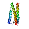
| ||||||||
|---|---|---|---|---|---|---|---|---|---|
| 1 |
| ||||||||
| Unit cell |
|
-Components
| #1: Protein | Mass: 11582.222 Da / Num. of mol.: 1 Source method: isolated from a genetically manipulated source Source: (gene. exp.) Staphylococcus aureus (bacteria) / Production host: Escherichia coli (E. coli) / References: UniProt: Q9F0J8, UniProt: A0A2S6DEV9*PLUS |
|---|---|
| #2: Water | ChemComp-HOH / Water Mass: 18.015 Da / Num. of mol.: 19 / Source method: isolated from a natural source / Formula: H2O Mass: 18.015 Da / Num. of mol.: 19 / Source method: isolated from a natural source / Formula: H2O |
-Experimental details
-Experiment
| Experiment | Method: X-RAY DIFFRACTION / Number of used crystals: 1 |
|---|
- Sample preparation
| Crystal | Density Matthews: 2.98 Å3/Da / Density % sol: 58.76 % |
|---|---|
| Crystal grow | Temperature: 294.15 K / Method: vapor diffusion, sitting drop / Details: 40%PEG3350 0.1M Bis-Tris 0.2M Na-thiocyanate |
-Data collection
| Diffraction | Mean temperature: 100 K | |||||||||||||||||||||||||||||||||||||||||||||||||||||||||||||||||||||||||||||||||||||||||||||||||||
|---|---|---|---|---|---|---|---|---|---|---|---|---|---|---|---|---|---|---|---|---|---|---|---|---|---|---|---|---|---|---|---|---|---|---|---|---|---|---|---|---|---|---|---|---|---|---|---|---|---|---|---|---|---|---|---|---|---|---|---|---|---|---|---|---|---|---|---|---|---|---|---|---|---|---|---|---|---|---|---|---|---|---|---|---|---|---|---|---|---|---|---|---|---|---|---|---|---|---|---|---|
| Diffraction source | Source: SYNCHROTRON / Site: ALBA / Beamline: XALOC / Wavelength: 0.97926 Å | |||||||||||||||||||||||||||||||||||||||||||||||||||||||||||||||||||||||||||||||||||||||||||||||||||
| Detector | Type: DECTRIS PILATUS3 S 6M / Detector: PIXEL / Date: Jun 8, 2017 | |||||||||||||||||||||||||||||||||||||||||||||||||||||||||||||||||||||||||||||||||||||||||||||||||||
| Radiation | Protocol: SINGLE WAVELENGTH / Monochromatic (M) / Laue (L): M / Scattering type: x-ray | |||||||||||||||||||||||||||||||||||||||||||||||||||||||||||||||||||||||||||||||||||||||||||||||||||
| Radiation wavelength | Wavelength: 0.97926 Å / Relative weight: 1 | |||||||||||||||||||||||||||||||||||||||||||||||||||||||||||||||||||||||||||||||||||||||||||||||||||
| Reflection | Resolution: 2.2→66.993 Å / Num. obs: 6522 / % possible obs: 96.7 % / Redundancy: 18.4 % / CC1/2: 0.999 / Rpim(I) all: 0.027 / Rrim(I) all: 0.116 / Rsym value: 0.112 / Net I/av σ(I): 3.5 / Net I/σ(I): 14.4 | |||||||||||||||||||||||||||||||||||||||||||||||||||||||||||||||||||||||||||||||||||||||||||||||||||
| Reflection shell | Diffraction-ID: 1
|
-Phasing
| Phasing | Method: SAD |
|---|
- Processing
| Software |
| ||||||||||||||||||||||||||||||||||||||||||||||||||||||||||||
|---|---|---|---|---|---|---|---|---|---|---|---|---|---|---|---|---|---|---|---|---|---|---|---|---|---|---|---|---|---|---|---|---|---|---|---|---|---|---|---|---|---|---|---|---|---|---|---|---|---|---|---|---|---|---|---|---|---|---|---|---|---|
| Refinement | Method to determine structure: SAD / Resolution: 2.2→66.99 Å / Cor.coef. Fo:Fc: 0.942 / Cor.coef. Fo:Fc free: 0.943 / SU R Cruickshank DPI: 0.2484 / Cross valid method: THROUGHOUT / σ(F): 0 / ESU R: 0.248 / ESU R Free: 0.198 Details: HYDROGENS HAVE BEEN ADDED IN THE RIDING POSITIONS U VALUES : REFINED INDIVIDUALLY
| ||||||||||||||||||||||||||||||||||||||||||||||||||||||||||||
| Solvent computation | Ion probe radii: 0.8 Å / Shrinkage radii: 0.8 Å / VDW probe radii: 1.2 Å | ||||||||||||||||||||||||||||||||||||||||||||||||||||||||||||
| Displacement parameters | Biso max: 149.85 Å2 / Biso mean: 67.634 Å2 / Biso min: 31.37 Å2
| ||||||||||||||||||||||||||||||||||||||||||||||||||||||||||||
| Refinement step | Cycle: final / Resolution: 2.2→66.99 Å
| ||||||||||||||||||||||||||||||||||||||||||||||||||||||||||||
| Refine LS restraints |
| ||||||||||||||||||||||||||||||||||||||||||||||||||||||||||||
| LS refinement shell | Resolution: 2.2→2.257 Å / Rfactor Rfree error: 0 / Total num. of bins used: 20
|
