 Movie
Movie Controller

[English] 日本語
 Yorodumi
- PDB-1row: Structure of SSP-19, an MSP-domain protein like family member in ... -
+ Open data

- Basic information
| Entry | Database: PDB / ID: 1row | ||||||
|---|---|---|---|---|---|---|---|
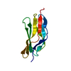
| Title | Structure of SSP-19, an MSP-domain protein like family member in Caenorhabditis elegans | ||||||
 Components Components | MSP-domain protein like family member | ||||||
 Keywords Keywords |  STRUCTURAL PROTEIN / BETA BARREL / Structural Genomics / PSI / Protein Structure Initiative / Southeast Collaboratory for Structural Genomics / SECSG STRUCTURAL PROTEIN / BETA BARREL / Structural Genomics / PSI / Protein Structure Initiative / Southeast Collaboratory for Structural Genomics / SECSG | ||||||
| Function / homology |  Function and homology information Function and homology information | ||||||
| Biological species |  Caenorhabditis elegans (invertebrata) Caenorhabditis elegans (invertebrata) | ||||||
| Method | X-RAY DIFFRACTION / MOLECULAR REPLACEMENT / Resolution: 2 Å | ||||||
 Authors Authors | Schormann, N. / Symersky, J. / Carson, M. / Luo, M. / Lin, G. / Li, S. / Qiu, S. / Arabashi, A. / Bunzel, B. / Luo, D. ...Schormann, N. / Symersky, J. / Carson, M. / Luo, M. / Lin, G. / Li, S. / Qiu, S. / Arabashi, A. / Bunzel, B. / Luo, D. / Nagy, L. / Gray, R. / Luan, C.-H. / Zhang, J. / Lu, S. / DeLucas, L. / Southeast Collaboratory for Structural Genomics (SECSG) | ||||||
 Citation Citation | Journal: Acta Crystallogr.,Sect.D / Year: 2004 Title: Structure of sperm-specific protein SSP-19 from Caenorhabditis elegans. Authors: Schormann, N. / Symersky, J. / Luo, M. | ||||||
| History |
|
- Structure visualization
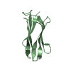
| Structure viewer | Molecule: MolmilJmol/JSmol |
|---|
- Downloads & links
-Download
| PDBx/mmCIF format | 1row.cif.gz | 53.3 KB | Display | PDBx/mmCIF format |
|---|---|---|---|---|
| PDB format | pdb1row.ent.gz | 37.9 KB | Display | PDB format |
| PDBx/mmJSON format | 1row.json.gz | Tree view | PDBx/mmJSON format | |
| Others |  Other downloads Other downloads |
-Validation report
| Arichive directory | https://data.pdbj.org/pub/pdb/validation_reports/ro/1rowftp://data.pdbj.org/pub/pdb/validation_reports/ro/1row | HTTPS FTP |
|---|
-Related structure data
| Related structure data |  1m1sS S: Starting model for refinement |
|---|---|
| Similar structure data | |
| Other databases |


-Links
 PDBj
PDBj- Assembly
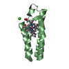
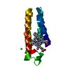
| Deposited unit | 
| ||||||||||
|---|---|---|---|---|---|---|---|---|---|---|---|
| 1 | 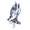
| ||||||||||
| 2 | 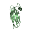
| ||||||||||
| Unit cell |
| ||||||||||
| Details | The biological unit is a monomer |
-Components
| #1: Protein | Mass: 11022.486 Da / Num. of mol.: 2 Source method: isolated from a genetically manipulated source Source: (gene. exp.) Caenorhabditis elegans (invertebrata) / Strain: BRISTOL N2 (Clone C55C2) / Cell: SPERM / Gene: SSP-19 / Plasmid: pET28B / Species (production host): Escherichia coli / Production host:  Escherichia coli BL21(DE3) (bacteria) / Strain (production host): BL21(DE3) / References: UniProt: O01829 Escherichia coli BL21(DE3) (bacteria) / Strain (production host): BL21(DE3) / References: UniProt: O01829#2: Water | ChemComp-HOH / | Water Mass: 18.015 Da / Num. of mol.: 157 / Source method: isolated from a natural source / Formula: H2O Mass: 18.015 Da / Num. of mol.: 157 / Source method: isolated from a natural source / Formula: H2O |
|---|
-Experimental details
-Experiment
| Experiment | Method: X-RAY DIFFRACTION / Number of used crystals: 1 |
|---|
- Sample preparation
| Crystal | Density Matthews: 1.96 Å3/Da / Density % sol: 36.8 % | |||||||||||||||||||||||||||||||||||||||||||||||||
|---|---|---|---|---|---|---|---|---|---|---|---|---|---|---|---|---|---|---|---|---|---|---|---|---|---|---|---|---|---|---|---|---|---|---|---|---|---|---|---|---|---|---|---|---|---|---|---|---|---|---|
| Crystal grow | Temperature: 295 K / Method: vapor diffusion, hanging drop / pH: 5.6 Details: 1.7M lithium sulfate, 0.05M Mes, 0.1M magnesium chloride, pH 5.6, VAPOR DIFFUSION, HANGING DROP, temperature 295K | |||||||||||||||||||||||||||||||||||||||||||||||||
| Crystal grow | *PLUS Temperature: 295 K / pH: 7.5 / Method: vapor diffusion, hanging drop | |||||||||||||||||||||||||||||||||||||||||||||||||
| Components of the solutions | *PLUS
|
-Data collection
| Diffraction | Mean temperature: 100 K |
|---|---|
| Diffraction source | Source: ROTATING ANODE / Type: RIGAKU RU200 / Wavelength: 1.5418 Å |
| Detector | Type: RIGAKU RAXIS IV / Detector: IMAGE PLATE / Date: Oct 24, 2003 / Details: OSMIC MIRRORS |
| Radiation | Monochromator: Graphite / Protocol: SINGLE WAVELENGTH / Monochromatic (M) / Laue (L): M / Scattering type: x-ray |
| Radiation wavelength | Wavelength: 1.5418 Å / Relative weight: 1 |
| Reflection | Resolution: 2→50 Å / Num. all: 11037 / Num. obs: 11037 / % possible obs: 88.2 % / Observed criterion σ(I): 0.5 / Redundancy: 3.3 % / Biso Wilson estimate: 25.3 Å2 / Rmerge(I) obs: 0.048 / Rsym value: 0.048 / Net I/σ(I): 33.9 |
| Reflection shell | Resolution: 2→2.07 Å / Redundancy: 2.5 % / Rmerge(I) obs: 0.066 / Num. unique all: 747 / Rsym value: 0.066 / % possible all: 60.5 |
| Reflection | *PLUS Highest resolution: 2.2 Å / Num. obs: 8710 / % possible obs: 92.4 % / Num. measured all: 28027 / Rmerge(I) obs: 0.053 |
| Reflection shell | *PLUS Highest resolution: 2.2 Å / Lowest resolution: 2.28 Å / % possible obs: 89.7 % / Redundancy: 3 % / Num. unique obs: 852 / Rmerge(I) obs: 0.087 / Mean I/σ(I) obs: 21.9 |
- Processing
| Software |
| ||||||||||||||||||||||||||||||||||||
|---|---|---|---|---|---|---|---|---|---|---|---|---|---|---|---|---|---|---|---|---|---|---|---|---|---|---|---|---|---|---|---|---|---|---|---|---|---|
| Refinement | Method to determine structure: MOLECULAR REPLACEMENT Starting model: Homology model (Swiss Model)based on PDB Entry 1M1S Resolution: 2→19.56 Å / Rfactor Rfree error: 0.012 / Data cutoff high absF: 525587.12 / Data cutoff low absF: 0 / Isotropic thermal model: RESTRAINED / Cross valid method: THROUGHOUT / σ(F): 1 / Stereochemistry target values: Engh & Huber Details: In initial stages restrained NCS was used, but later the two monomers were refined independently
| ||||||||||||||||||||||||||||||||||||
| Solvent computation | Solvent model: FLAT MODEL / Bsol: 53.6843 Å2 / ksol: 0.363445 e/Å3 | ||||||||||||||||||||||||||||||||||||
| Displacement parameters | Biso mean: 26.9 Å2
| ||||||||||||||||||||||||||||||||||||
| Refine analyze |
| ||||||||||||||||||||||||||||||||||||
| Refinement step | Cycle: LAST / Resolution: 2→19.56 Å
| ||||||||||||||||||||||||||||||||||||
| Refine LS restraints |
| ||||||||||||||||||||||||||||||||||||
| LS refinement shell | Resolution: 2→2.13 Å / Rfactor Rfree error: 0.042 / Total num. of bins used: 6
| ||||||||||||||||||||||||||||||||||||
| Xplor file |
| ||||||||||||||||||||||||||||||||||||
| Refinement | *PLUS Highest resolution: 2.2 Å / Lowest resolution: 24.79 Å / Num. reflection obs: 8637 / % reflection Rfree: 5 % / Rfactor obs: 0.219 / Rfactor Rfree: 0.28 / Rfactor Rwork: 0.224 | ||||||||||||||||||||||||||||||||||||
| Solvent computation | *PLUS | ||||||||||||||||||||||||||||||||||||
| Displacement parameters | *PLUS | ||||||||||||||||||||||||||||||||||||
| Refine LS restraints | *PLUS
| ||||||||||||||||||||||||||||||||||||
| LS refinement shell | *PLUS Highest resolution: 2.2 Å / Lowest resolution: 2.34 Å / Rfactor Rfree: 0.327 / Rfactor Rwork: 0.207 / Num. reflection Rwork: 1217 / Rfactor obs: 0.237 |
