 Movie
Movie Controller

[English] 日本語
 Yorodumi
- PDB-6gyh: Crystal structure of the light-driven proton pump Coccomyxa subel... -
+ Open data

- Basic information
| Entry | Database: PDB / ID: 6gyh | |||||||||
|---|---|---|---|---|---|---|---|---|---|---|
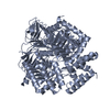
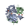
| Title | Crystal structure of the light-driven proton pump Coccomyxa subellipsoidea Rhodopsin CsR | |||||||||
 Components Components | Family A G protein-coupled receptor-like protein | |||||||||
 Keywords Keywords |  PROTON TRANSPORT / RETINAL / MICROBIAL-TYPE RHODOPSIN / LIGHT-DRIVEN PROTON PUMP / SEVEN TRANS-MEMBRANE / TRANSPORT PROTEIN / MEMBRANE PROTEIN PROTON TRANSPORT / RETINAL / MICROBIAL-TYPE RHODOPSIN / LIGHT-DRIVEN PROTON PUMP / SEVEN TRANS-MEMBRANE / TRANSPORT PROTEIN / MEMBRANE PROTEIN | |||||||||
| Function / homology |  Function and homology information: / photoreceptor activity / phototransduction / monoatomic ion channel activity / membrane => GO:0016020 Function and homology information: / photoreceptor activity / phototransduction / monoatomic ion channel activity / membrane => GO:0016020Similarity search - Function | |||||||||
| Biological species |  Coccomyxa subellipsoidea C-169 (plant) Coccomyxa subellipsoidea C-169 (plant) | |||||||||
| Method | X-RAY DIFFRACTION / SYNCHROTRON / MOLECULAR REPLACEMENT / Resolution: 2 Å | |||||||||
 Authors Authors | Szczepek, M. / Schmidt, A. / Scheerer, P. | |||||||||
| Funding support |  Germany, 2items Germany, 2items
| |||||||||
 Citation Citation | Journal: Sci.Signal. / Year: 2019 Title: Design of a light-gated proton channel based on the crystal structure ofCoccomyxarhodopsin. Authors: Fudim, R. / Szczepek, M. / Vierock, J. / Vogt, A. / Schmidt, A. / Kleinau, G. / Fischer, P. / Bartl, F. / Scheerer, P. / Hegemann, P. | |||||||||
| History |
|
- Structure visualization
| Structure viewer | Molecule: MolmilJmol/JSmol |
|---|
- Downloads & links
-Download
| PDBx/mmCIF format | 6gyh.cif.gz | 109.3 KB | Display | PDBx/mmCIF format |
|---|---|---|---|---|
| PDB format | pdb6gyh.ent.gz | 82.5 KB | Display | PDB format |
| PDBx/mmJSON format | 6gyh.json.gz | Tree view | PDBx/mmJSON format | |
| Others |  Other downloads Other downloads |
-Validation report
| Arichive directory | https://data.pdbj.org/pub/pdb/validation_reports/gy/6gyhftp://data.pdbj.org/pub/pdb/validation_reports/gy/6gyh | HTTPS FTP |
|---|
-Related structure data
| Related structure data | 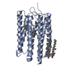 3am6S S: Starting model for refinement |
|---|---|
| Similar structure data |

-Links
 PDBj
PDBj


- Assembly
| Deposited unit | 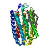
| ||||||||
|---|---|---|---|---|---|---|---|---|---|
| 1 | 
| ||||||||
| Unit cell |
|
-Components
| #1: Protein | Mass: 25733.086 Da / Num. of mol.: 1 Source method: isolated from a genetically manipulated source Source: (gene. exp.) Coccomyxa subellipsoidea C-169 (plant) / Gene: COCSUDRAFT_42526 / Production host:   Spodoptera frugiperda (fall armyworm) / References: UniProt: I0YUS5 Spodoptera frugiperda (fall armyworm) / References: UniProt: I0YUS5 | ||
|---|---|---|---|
| #2: Chemical | ChemComp-RET / Retinal  Mass: 284.436 Da / Num. of mol.: 1 / Source method: obtained synthetically / Formula: C20H28O Mass: 284.436 Da / Num. of mol.: 1 / Source method: obtained synthetically / Formula: C20H28O | ||
| #3: Chemical | ChemComp-CLR / Cholesterol  Mass: 386.654 Da / Num. of mol.: 1 / Source method: obtained synthetically / Formula: C27H46O Mass: 386.654 Da / Num. of mol.: 1 / Source method: obtained synthetically / Formula: C27H46O | ||
| #4: Chemical | ChemComp-OLB / (   Mass: 356.540 Da / Num. of mol.: 4 / Source method: obtained synthetically / Formula: C21H40O4 Mass: 356.540 Da / Num. of mol.: 4 / Source method: obtained synthetically / Formula: C21H40O4#5: Water | ChemComp-HOH / | Water Mass: 18.015 Da / Num. of mol.: 42 / Source method: isolated from a natural source / Formula: H2O Mass: 18.015 Da / Num. of mol.: 42 / Source method: isolated from a natural source / Formula: H2O |
-Experimental details
-Experiment
| Experiment | Method: X-RAY DIFFRACTION / Number of used crystals: 1 |
|---|
- Sample preparation
| Crystal | Density Matthews: 2.62 Å3/Da / Density % sol: 53.05 % |
|---|---|
| Crystal grow | Temperature: 293 K / Method: vapor diffusion Details: 1-(9Z-octadecenoyl)-rac-glycerol, 10 % cholesterol, 42 % v/v polyethylene glycol 400; 100 mM MES; pH 6.5, 150 mM sodium acetate |
-Data collection
| Diffraction | Mean temperature: 100 K |
|---|---|
| Diffraction source | Source: SYNCHROTRON / Site: ESRF  / Beamline: ID23-2 / Wavelength: 0.8729 Å / Beamline: ID23-2 / Wavelength: 0.8729 Å |
| Detector | Type: DECTRIS PILATUS3 2M / Detector: PIXEL / Date: Oct 4, 2016 |
| Radiation | Protocol: SINGLE WAVELENGTH / Monochromatic (M) / Laue (L): M / Scattering type: x-ray |
| Radiation wavelength | Wavelength: 0.8729 Å / Relative weight: 1 |
| Reflection | Resolution: 2→32.91 Å / Num. obs: 22100 / % possible obs: 100 % / Redundancy: 4.4 % / CC1/2: 0.997 / Rpim(I) all: 0.066 / Net I/σ(I): 6.8 |
| Reflection shell | Resolution: 2→2.05 Å / Redundancy: 4.6 % / Mean I/σ(I) obs: 0.8 / CC1/2: 0.357 / Rpim(I) all: 0.901 / % possible all: 100 |
- Processing
| Software |
| ||||||||||||||||||||||||||||||||||||||||||||||||||||||||||||||||||||||||||||||||||||||||||||||||||||||||||||||||||||||||||||||||||||||||||||||||||||||||||||||||||||||||||||||||||||||
|---|---|---|---|---|---|---|---|---|---|---|---|---|---|---|---|---|---|---|---|---|---|---|---|---|---|---|---|---|---|---|---|---|---|---|---|---|---|---|---|---|---|---|---|---|---|---|---|---|---|---|---|---|---|---|---|---|---|---|---|---|---|---|---|---|---|---|---|---|---|---|---|---|---|---|---|---|---|---|---|---|---|---|---|---|---|---|---|---|---|---|---|---|---|---|---|---|---|---|---|---|---|---|---|---|---|---|---|---|---|---|---|---|---|---|---|---|---|---|---|---|---|---|---|---|---|---|---|---|---|---|---|---|---|---|---|---|---|---|---|---|---|---|---|---|---|---|---|---|---|---|---|---|---|---|---|---|---|---|---|---|---|---|---|---|---|---|---|---|---|---|---|---|---|---|---|---|---|---|---|---|---|---|---|
| Refinement | Method to determine structure: MOLECULAR REPLACEMENT Starting model: 3AM6 Resolution: 2→32.91 Å / Cor.coef. Fo:Fc: 0.956 / Cor.coef. Fo:Fc free: 0.919 / SU B: 11.792 / SU ML: 0.133 / Cross valid method: THROUGHOUT / ESU R: 0.266 / ESU R Free: 0.137 / Details: HYDROGENS HAVE BEEN ADDED IN THE RIDING POSITIONS
| ||||||||||||||||||||||||||||||||||||||||||||||||||||||||||||||||||||||||||||||||||||||||||||||||||||||||||||||||||||||||||||||||||||||||||||||||||||||||||||||||||||||||||||||||||||||
| Solvent computation | Ion probe radii: 0.8 Å / Shrinkage radii: 0.8 Å / VDW probe radii: 1.2 Å | ||||||||||||||||||||||||||||||||||||||||||||||||||||||||||||||||||||||||||||||||||||||||||||||||||||||||||||||||||||||||||||||||||||||||||||||||||||||||||||||||||||||||||||||||||||||
| Displacement parameters | Biso mean: 43.432 Å2
| ||||||||||||||||||||||||||||||||||||||||||||||||||||||||||||||||||||||||||||||||||||||||||||||||||||||||||||||||||||||||||||||||||||||||||||||||||||||||||||||||||||||||||||||||||||||
| Refinement step | Cycle: 1 / Resolution: 2→32.91 Å
| ||||||||||||||||||||||||||||||||||||||||||||||||||||||||||||||||||||||||||||||||||||||||||||||||||||||||||||||||||||||||||||||||||||||||||||||||||||||||||||||||||||||||||||||||||||||
| Refine LS restraints |
|