| Entry | Database: PDB / ID: 6fwb
|
|---|
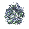
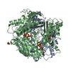
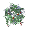
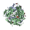
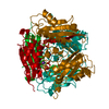
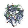
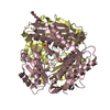
| Title | Crystal structure of Mat2A at 1.79 Angstron resolution |
|---|
 Components Components | S-adenosylmethionine synthase isoform type-2 |
|---|
 Keywords Keywords |  HYDROLASE / S-adenosylmethionine synthetase 2 HYDROLASE / S-adenosylmethionine synthetase 2 |
|---|
| Function / homology |  Function and homology information Function and homology information
methionine adenosyltransferase complex / methionine adenosyltransferase / methionine adenosyltransferase activity / S-adenosylmethionine biosynthetic process / Methylation / protein heterooligomerization / protein hexamerization / small molecule binding / cellular response to leukemia inhibitory factor / one-carbon metabolic process ...methionine adenosyltransferase complex / methionine adenosyltransferase / methionine adenosyltransferase activity / S-adenosylmethionine biosynthetic process / Methylation / protein heterooligomerization / protein hexamerization / small molecule binding / cellular response to leukemia inhibitory factor / one-carbon metabolic process / ATP binding / identical protein binding / metal ion binding / cytosolSimilarity search - Function GMP Synthetase; Chain A, domain 3 - #10 / S-adenosylmethionine synthetase / S-adenosylmethionine synthetase, N-terminal / S-adenosylmethionine synthetase, central domain / S-adenosylmethionine synthetase, C-terminal / S-adenosylmethionine synthetase, conserved site / S-adenosylmethionine synthetase superfamily / S-adenosylmethionine synthetase, N-terminal domain / S-adenosylmethionine synthetase, central domain / S-adenosylmethionine synthetase, C-terminal domain ...GMP Synthetase; Chain A, domain 3 - #10 / S-adenosylmethionine synthetase / S-adenosylmethionine synthetase, N-terminal / S-adenosylmethionine synthetase, central domain / S-adenosylmethionine synthetase, C-terminal / S-adenosylmethionine synthetase, conserved site / S-adenosylmethionine synthetase superfamily / S-adenosylmethionine synthetase, N-terminal domain / S-adenosylmethionine synthetase, central domain / S-adenosylmethionine synthetase, C-terminal domain / S-adenosylmethionine synthase signature 1. / S-adenosylmethionine synthase signature 2. / GMP Synthetase; Chain A, domain 3 / 2-Layer Sandwich / Alpha BetaSimilarity search - Domain/homology |
|---|
| Biological species |  Homo sapiens (human) Homo sapiens (human) |
|---|
| Method | X-RAY DIFFRACTION / SYNCHROTRON / MOLECULAR REPLACEMENT / Resolution: 1.79 Å |
|---|
 Authors Authors | Zhou, A. / Wei, Z. / Bai, J. / Wang, H. |
|---|
 Citation Citation | Journal: Ebiomedicine / Year: 2019
Title: Identification of a natural inhibitor of methionine adenosyltransferase 2A regulating one-carbon metabolism in keratinocytes.
Authors: Bai, J. / Gao, Y. / Chen, L. / Yin, Q. / Lou, F. / Wang, Z. / Xu, Z. / Zhou, H. / Li, Q. / Cai, W. / Sun, Y. / Niu, L. / Wang, H. / Wei, Z. / Lu, S. / Zhou, A. / Zhang, J. / Wang, H. |
|---|
| History | | Deposition | Mar 6, 2018 | Deposition site: PDBE / Processing site: PDBE |
|---|
| Revision 1.0 | Mar 27, 2019 | Provider: repository / Type: Initial release |
|---|
| Revision 1.1 | Jun 12, 2019 | Group: Data collection / Structure summary
Category: audit_author / database_PDB_rev / database_PDB_rev_record
Item: _audit_author.name |
|---|
| Revision 1.2 | May 8, 2024 | Group: Data collection / Database references / Refinement description
Category: chem_comp_atom / chem_comp_bond ...chem_comp_atom / chem_comp_bond / database_2 / struct_ncs_dom_lim
Item: _database_2.pdbx_DOI / _database_2.pdbx_database_accession ..._database_2.pdbx_DOI / _database_2.pdbx_database_accession / _struct_ncs_dom_lim.beg_auth_comp_id / _struct_ncs_dom_lim.beg_label_asym_id / _struct_ncs_dom_lim.beg_label_comp_id / _struct_ncs_dom_lim.beg_label_seq_id / _struct_ncs_dom_lim.end_auth_comp_id / _struct_ncs_dom_lim.end_label_asym_id / _struct_ncs_dom_lim.end_label_comp_id / _struct_ncs_dom_lim.end_label_seq_id |
|---|
|
|---|
 Movie
Movie Controller


 Open data
 Basic information
 Structure visualization
 Downloads & links
 Other downloads
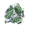


 PDBj
PDBj Assembly


 Mass: 96.063 Da / Num. of mol.: 19 / Source method: obtained synthetically / Formula: SO4
 Mass: 194.226 Da / Num. of mol.: 7 / Source method: obtained synthetically / Formula: C8H18O5 / Comment: precipitant*YM
 Mass: 22.990 Da / Num. of mol.: 1 / Source method: obtained synthetically / Formula: Na
 Mass: 92.094 Da / Num. of mol.: 2 / Source method: obtained synthetically / Formula: C3H8O3
 Sample preparation
 / Beamline: BL17U1 / Wavelength: 1.07822 Å
 Processing