 Movie
Movie Controller


+ Open data

- Basic information
| Entry | Database: PDB / ID: 6gu2 | |||||||||
|---|---|---|---|---|---|---|---|---|---|---|
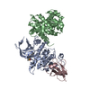
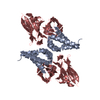
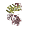
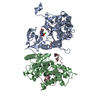
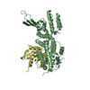
| Title | CDK1/CyclinB/Cks2 in complex with Flavopiridol | |||||||||
 Components Components |
| |||||||||
 Keywords Keywords |  CELL CYCLE / CDK1 / CYCLINB1 / CKS2 / INHIBITOR CELL CYCLE / CDK1 / CYCLINB1 / CKS2 / INHIBITOR | |||||||||
| Function / homology |  Function and homology information Function and homology informationregulation of Schwann cell differentiation / pronuclear fusion / cyclin B1-CDK1 complex / positive regulation of mitochondrial ATP synthesis coupled electron transport / Mitotic Prophase / positive regulation of mitotic sister chromatid segregation / histone kinase activity / Golgi disassembly / microtubule cytoskeleton organization involved in mitosis / G2/M DNA replication checkpoint ...regulation of Schwann cell differentiation / pronuclear fusion / cyclin B1-CDK1 complex / positive regulation of mitochondrial ATP synthesis coupled electron transport / Mitotic Prophase / positive regulation of mitotic sister chromatid segregation / histone kinase activity / Golgi disassembly / microtubule cytoskeleton organization involved in mitosis / G2/M DNA replication checkpoint / E2F-enabled inhibition of pre-replication complex formation / ventricular cardiac muscle cell development / Depolymerization of the Nuclear Lamina / positive regulation of attachment of spindle microtubules to kinetochore / MASTL Facilitates Mitotic Progression / regulation of mitotic cell cycle spindle assembly checkpoint / Activation of NIMA Kinases NEK9, NEK6, NEK7 / Phosphorylation of Emi1 / Phosphorylation of proteins involved in the G2/M transition by Cyclin A:Cdc2 complexes / patched binding / cyclin A2-CDK1 complex / Transcriptional regulation by RUNX2 / Nuclear Pore Complex (NPC) Disassembly / Phosphorylation of the APC/C / outer kinetochore / meiosis I / mitotic cell cycle phase transition / Transcription of E2F targets under negative control by p107 (RBL1) and p130 (RBL2) in complex with HDAC1 / Initiation of Nuclear Envelope (NE) Reformation / protein localization to kinetochore / Polo-like kinase mediated events / Golgi Cisternae Pericentriolar Stack Reorganization / cyclin-dependent protein serine/threonine kinase activator activity / Condensation of Prometaphase Chromosomes / chromosome condensation / response to copper ion / centrosome cycle / [RNA-polymerase]-subunit kinase / cyclin-dependent protein serine/threonine kinase regulator activity / SCF ubiquitin ligase complex / mitotic metaphase chromosome alignment / G1/S-Specific Transcription / cyclin-dependent protein kinase activity / MAPK3 (ERK1) activation / response to amine / ubiquitin-like protein ligase binding / mitotic G2 DNA damage checkpoint signaling / Regulation of APC/C activators between G1/S and early anaphase / regulation of embryonic development / cellular response to organic cyclic compound / cyclin-dependent protein kinase holoenzyme complex / response to axon injury / cyclin-dependent kinase / animal organ regeneration / cyclin-dependent protein serine/threonine kinase activity / response to cadmium ion / Chk1/Chk2(Cds1) mediated inactivation of Cyclin B:Cdk1 complex / Cyclin A/B1/B2 associated events during G2/M transition / positive regulation of cardiac muscle cell proliferation / Loss of Nlp from mitotic centrosomes / Loss of proteins required for interphase microtubule organization from the centrosome / Recruitment of mitotic centrosome proteins and complexes / ERK1 and ERK2 cascade / Hsp70 protein binding / Resolution of Sister Chromatid Cohesion / Recruitment of NuMA to mitotic centrosomes / epithelial cell differentiation / APC/C:Cdc20 mediated degradation of Cyclin B / regulation of mitotic cell cycle / Anchoring of the basal body to the plasma membrane / positive regulation of G2/M transition of mitotic cell cycle / cyclin binding / positive regulation of mitotic cell cycle / RNA polymerase II CTD heptapeptide repeat kinase activity / AURKA Activation by TPX2 / mitotic spindle organization / TP53 Regulates Transcription of Genes Involved in G2 Cell Cycle Arrest / Condensation of Prophase Chromosomes / response to activity / positive regulation of DNA replication / ubiquitin binding / spindle microtubule / Cdc20:Phospho-APC/C mediated degradation of Cyclin A / G1/S transition of mitotic cell cycle / peptidyl-threonine phosphorylation / MAPK6/MAPK4 signaling / PKR-mediated signaling / regulation of circadian rhythm / mitotic spindle / microtubule cytoskeleton organization / response to toxic substance / cellular response to hydrogen peroxide / spindle pole / positive regulation of protein import into nucleus / positive regulation of protein localization to nucleus / The role of GTSE1 in G2/M progression after G2 checkpoint / rhythmic process / G2/M transition of mitotic cell cycle / positive regulation of fibroblast proliferation / Regulation of PLK1 Activity at G2/M TransitionSimilarity search - Function | |||||||||
| Biological species |  Homo sapiens (human) Homo sapiens (human) | |||||||||
| Method | X-RAY DIFFRACTION / SYNCHROTRON / MOLECULAR REPLACEMENT / Resolution: 2 Å | |||||||||
 Authors Authors | Wood, D.J. / Korolchuk, S. / Tatum, N.J. / Wang, L.Z. / Endicott, J.A. / Noble, M.E.M. / Martin, M.P. | |||||||||
| Funding support |  United Kingdom, 2items United Kingdom, 2items
| |||||||||
 Citation Citation | Journal: Cell Chem Biol / Year: 2019 Title: Differences in the Conformational Energy Landscape of CDK1 and CDK2 Suggest a Mechanism for Achieving Selective CDK Inhibition. Authors: Wood, D.J. / Korolchuk, S. / Tatum, N.J. / Wang, L.Z. / Endicott, J.A. / Noble, M.E.M. / Martin, M.P. | |||||||||
| History |
|
- Structure visualization
| Structure viewer | Molecule: MolmilJmol/JSmol |
|---|
- Downloads & links
-Download
| PDBx/mmCIF format | 6gu2.cif.gz | 147.4 KB | Display | PDBx/mmCIF format |
|---|---|---|---|---|
| PDB format | pdb6gu2.ent.gz | 113.1 KB | Display | PDB format |
| PDBx/mmJSON format | 6gu2.json.gz | Tree view | PDBx/mmJSON format | |
| Others |  Other downloads Other downloads |
-Validation report
| Arichive directory | https://data.pdbj.org/pub/pdb/validation_reports/gu/6gu2ftp://data.pdbj.org/pub/pdb/validation_reports/gu/6gu2 | HTTPS FTP |
|---|
-Related structure data
| Related structure data |  6gu3C 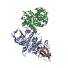 6gu4C 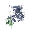 6gu6C  6gu7C  6gubC 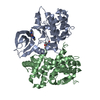 6gucC 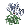 6gueC  6gufC  6guhC  6gukC  4y72S S: Starting model for refinement C: citing same article ( |
|---|---|
| Similar structure data |


-Links
 PDBj
PDBj


- Assembly
| Deposited unit | 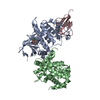
| ||||||||
|---|---|---|---|---|---|---|---|---|---|
| 1 |
| ||||||||
| Unit cell |
|
-Components
| #1: Protein | / CDK1 / Cell division control protein 2 homolog / Cell division protein kinase 1 / p34 protein kinase Mass: 34553.883 Da / Num. of mol.: 1 Source method: isolated from a genetically manipulated source Source: (gene. exp.) Homo sapiens (human) / Gene: CDK1, CDC2, CDC28A, CDKN1, P34CDC2 / Plasmid: pVL1393 / Cell line (production host): SF9 / Production host:   Spodoptera frugiperda (fall armyworm) Spodoptera frugiperda (fall armyworm)References: UniProt: P06493, cyclin-dependent kinase, [RNA-polymerase]-subunit kinase |
|---|---|
| #2: Protein | Mass: 31369.682 Da / Num. of mol.: 1 Source method: isolated from a genetically manipulated source Source: (gene. exp.) Homo sapiens (human) / Gene: CCNB1, CCNB / Plasmid: pET28A / Production host:  Escherichia coli BL21(DE3) (bacteria) / References: UniProt: P14635 Escherichia coli BL21(DE3) (bacteria) / References: UniProt: P14635 |
| #3: Protein | Mass: 10290.819 Da / Num. of mol.: 1 Source method: isolated from a genetically manipulated source Source: (gene. exp.) Homo sapiens (human) / Gene: CKS2 / Production host: Escherichia coli (E. coli) / References: UniProt: P33552, UniProt: K9J4F7*PLUS |
| #4: Chemical | ChemComp-F9Z /   Mass: 401.840 Da / Num. of mol.: 1 / Source method: obtained synthetically / Formula: C21H20ClNO5 / Feature type: SUBJECT OF INVESTIGATION Mass: 401.840 Da / Num. of mol.: 1 / Source method: obtained synthetically / Formula: C21H20ClNO5 / Feature type: SUBJECT OF INVESTIGATION |
| #5: Water | ChemComp-HOH / Water Mass: 18.015 Da / Num. of mol.: 194 / Source method: isolated from a natural source / Formula: H2O Mass: 18.015 Da / Num. of mol.: 194 / Source method: isolated from a natural source / Formula: H2O |
-Experimental details
-Experiment
| Experiment | Method: X-RAY DIFFRACTION / Number of used crystals: 1 |
|---|
- Sample preparation
| Crystal | Density Matthews: 2.46 Å3/Da / Density % sol: 49.96 % |
|---|---|
| Crystal grow | Temperature: 277 K / Method: vapor diffusion, sitting drop Details: 0.1M MES/IMIDAZOLE BUFFER (PH6.7), 6.5% MPD, 5% PEG4K, 10% PEG1K PROTEIN AT 10-12 MG/ML + 0.5mM Inhibitor |
-Data collection
| Diffraction | Mean temperature: 100 K |
|---|---|
| Diffraction source | Source: SYNCHROTRON / Site: Diamond / Beamline: I04 / Wavelength: 0.987 Å |
| Detector | Type: DECTRIS PILATUS 6M / Detector: PIXEL / Date: Jun 25, 2016 |
| Radiation | Protocol: SINGLE WAVELENGTH / Monochromatic (M) / Laue (L): M / Scattering type: x-ray |
| Radiation wavelength | Wavelength: 0.987 Å / Relative weight: 1 |
| Reflection | Resolution: 2→89.17 Å / Num. obs: 50412 / % possible obs: 100 % / Redundancy: 3.7 % / Rmerge(I) obs: 0.09 / Net I/σ(I): 6.9 |
| Reflection shell | Resolution: 2→2.05 Å / Redundancy: 3.7 % / Rmerge(I) obs: 1.01 / Mean I/σ(I) obs: 1.1 / % possible all: 100 |
- Processing
| Software |
| ||||||||||||||||||||||||||||||||||||||||||||||||||||||||||||||||||||||||||||||||||||||||||||||||||||||||||||||||||||||||||||||||||||||||||||||||||||||||||||||||||||||||||||||||||||||
|---|---|---|---|---|---|---|---|---|---|---|---|---|---|---|---|---|---|---|---|---|---|---|---|---|---|---|---|---|---|---|---|---|---|---|---|---|---|---|---|---|---|---|---|---|---|---|---|---|---|---|---|---|---|---|---|---|---|---|---|---|---|---|---|---|---|---|---|---|---|---|---|---|---|---|---|---|---|---|---|---|---|---|---|---|---|---|---|---|---|---|---|---|---|---|---|---|---|---|---|---|---|---|---|---|---|---|---|---|---|---|---|---|---|---|---|---|---|---|---|---|---|---|---|---|---|---|---|---|---|---|---|---|---|---|---|---|---|---|---|---|---|---|---|---|---|---|---|---|---|---|---|---|---|---|---|---|---|---|---|---|---|---|---|---|---|---|---|---|---|---|---|---|---|---|---|---|---|---|---|---|---|---|---|
| Refinement | Method to determine structure: MOLECULAR REPLACEMENT Starting model: 4Y72 Resolution: 2→89.17 Å / Cor.coef. Fo:Fc: 0.966 / Cor.coef. Fo:Fc free: 0.948 / Cross valid method: THROUGHOUT / ESU R: 0.174 / ESU R Free: 0.163 / Details: HYDROGENS HAVE BEEN ADDED IN THE RIDING POSITIONS
| ||||||||||||||||||||||||||||||||||||||||||||||||||||||||||||||||||||||||||||||||||||||||||||||||||||||||||||||||||||||||||||||||||||||||||||||||||||||||||||||||||||||||||||||||||||||
| Solvent computation | Ion probe radii: 0.8 Å / Shrinkage radii: 0.8 Å / VDW probe radii: 1.2 Å | ||||||||||||||||||||||||||||||||||||||||||||||||||||||||||||||||||||||||||||||||||||||||||||||||||||||||||||||||||||||||||||||||||||||||||||||||||||||||||||||||||||||||||||||||||||||
| Displacement parameters | Biso mean: 47.989 Å2
| ||||||||||||||||||||||||||||||||||||||||||||||||||||||||||||||||||||||||||||||||||||||||||||||||||||||||||||||||||||||||||||||||||||||||||||||||||||||||||||||||||||||||||||||||||||||
| Refinement step | Cycle: 1 / Resolution: 2→89.17 Å
| ||||||||||||||||||||||||||||||||||||||||||||||||||||||||||||||||||||||||||||||||||||||||||||||||||||||||||||||||||||||||||||||||||||||||||||||||||||||||||||||||||||||||||||||||||||||
| Refine LS restraints |
|