 Movie
Movie Controller

[English] 日本語
 Yorodumi
- PDB-6g7l: Retinal isomerization in bacteriorhodopsin revealed by a femtosec... -
+ Open data

- Basic information
| Entry | Database: PDB / ID: 6g7l | |||||||||||||||
|---|---|---|---|---|---|---|---|---|---|---|---|---|---|---|---|---|
| Title | Retinal isomerization in bacteriorhodopsin revealed by a femtosecond X-ray laser: 8.3 ms state structure | |||||||||||||||
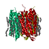
 Components Components | Bacteriorhodopsin | |||||||||||||||
 Keywords Keywords | PROTON TRANSPORT / retinal isomerization / ultrafast / femtosecond / room temperature / time resolved crystallography | |||||||||||||||
| Function / homology |  Function and homology informationphotoreceptor activity / phototransduction / proton transmembrane transport / monoatomic ion channel activity / plasma membrane Function and homology informationphotoreceptor activity / phototransduction / proton transmembrane transport / monoatomic ion channel activity / plasma membraneSimilarity search - Function | |||||||||||||||
| Biological species |  Halobacterium salinarum (Halophile) Halobacterium salinarum (Halophile) | |||||||||||||||
| Method | X-RAY DIFFRACTION / FREE ELECTRON LASER / MOLECULAR REPLACEMENT / Resolution: 1.9 Å | |||||||||||||||
 Authors Authors | Nogly, P. / Weinert, T. / James, D. / Cabajo, S. / Ozerov, D. / Furrer, A. / Gashi, D. / Borin, V. / Skopintsev, P. / Jaeger, K. ...Nogly, P. / Weinert, T. / James, D. / Cabajo, S. / Ozerov, D. / Furrer, A. / Gashi, D. / Borin, V. / Skopintsev, P. / Jaeger, K. / Nass, K. / Bath, P. / Bosman, R. / Koglin, J. / Seaberg, M. / Lane, T. / Kekilli, D. / Bruenle, S. / Tanaka, T. / Wu, W. / Milne, C. / White, T. / Barty, A. / Weierstall, U. / Panneels, V. / Nango, E. / Iwata, S. / Hunter, M. / Schapiro, I. / Schertler, G. / Neutze, R. / Standfuss, J. | |||||||||||||||
| Funding support |  Switzerland, 4items Switzerland, 4items
| |||||||||||||||
 Citation Citation | Journal: Science / Year: 2018 Title: Retinal isomerization in bacteriorhodopsin captured by a femtosecond x-ray laser. Authors: Nogly, P. / Weinert, T. / James, D. / Carbajo, S. / Ozerov, D. / Furrer, A. / Gashi, D. / Borin, V. / Skopintsev, P. / Jaeger, K. / Nass, K. / Bath, P. / Bosman, R. / Koglin, J. / Seaberg, M. ...Authors: Nogly, P. / Weinert, T. / James, D. / Carbajo, S. / Ozerov, D. / Furrer, A. / Gashi, D. / Borin, V. / Skopintsev, P. / Jaeger, K. / Nass, K. / Bath, P. / Bosman, R. / Koglin, J. / Seaberg, M. / Lane, T. / Kekilli, D. / Brunle, S. / Tanaka, T. / Wu, W. / Milne, C. / White, T. / Barty, A. / Weierstall, U. / Panneels, V. / Nango, E. / Iwata, S. / Hunter, M. / Schapiro, I. / Schertler, G. / Neutze, R. / Standfuss, J. | |||||||||||||||
| History |
|
- Structure visualization
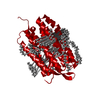
| Structure viewer | Molecule: MolmilJmol/JSmol |
|---|
- Downloads & links
-Download
| PDBx/mmCIF format | 6g7l.cif.gz | 110.3 KB | Display | PDBx/mmCIF format |
|---|---|---|---|---|
| PDB format | pdb6g7l.ent.gz | 84.7 KB | Display | PDB format |
| PDBx/mmJSON format | 6g7l.json.gz | Tree view | PDBx/mmJSON format | |
| Others |  Other downloads Other downloads |
-Validation report
| Arichive directory | https://data.pdbj.org/pub/pdb/validation_reports/g7/6g7lftp://data.pdbj.org/pub/pdb/validation_reports/g7/6g7l | HTTPS FTP |
|---|
-Related structure data
| Related structure data | 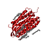 6g7hC 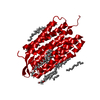 6g7iC  6g7jC 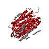 6g7kC  5j7aS S: Starting model for refinement C: citing same article ( |
|---|---|
| Similar structure data |


-Links
 PDBj
PDBj


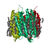
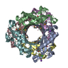
- Assembly
| Deposited unit | 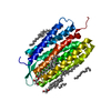
| ||||||||
|---|---|---|---|---|---|---|---|---|---|
| 1 | 
| ||||||||
| Unit cell |
| ||||||||
| Components on special symmetry positions |
|
-Components
| #1: Protein | / BR / Bacterioopsin / BO Mass: 28270.084 Da / Num. of mol.: 1 / Source method: isolated from a natural source Source: (natural) Halobacterium salinarum (strain ATCC 700922 / JCM 11081 / NRC-1) (Halophile)References: UniProt: P02945 | ||||
|---|---|---|---|---|---|
| #2: Chemical | ChemComp-RET / Retinal  Mass: 284.436 Da / Num. of mol.: 1 / Source method: obtained synthetically / Formula: C20H28O Mass: 284.436 Da / Num. of mol.: 1 / Source method: obtained synthetically / Formula: C20H28O | ||||
| #3: Chemical | ChemComp-LI1 /   Mass: 639.130 Da / Num. of mol.: 7 / Source method: obtained synthetically / Formula: C42H86O3 Mass: 639.130 Da / Num. of mol.: 7 / Source method: obtained synthetically / Formula: C42H86O3#4: Chemical | ChemComp-OLC / (   Mass: 356.540 Da / Num. of mol.: 4 / Source method: obtained synthetically / Formula: C21H40O4 Mass: 356.540 Da / Num. of mol.: 4 / Source method: obtained synthetically / Formula: C21H40O4#5: Water | ChemComp-HOH / | Water Mass: 18.015 Da / Num. of mol.: 59 / Source method: isolated from a natural source / Formula: H2O Mass: 18.015 Da / Num. of mol.: 59 / Source method: isolated from a natural source / Formula: H2O |
-Experimental details
-Experiment
| Experiment | Method: X-RAY DIFFRACTION / Number of used crystals: 1 |
|---|
- Sample preparation
| Crystal | Density Matthews: 2.2 Å3/Da / Density % sol: 44.17 % / Description: plates |
|---|---|
| Crystal grow | Temperature: 294 K / Method: lipidic cubic phase / pH: 5.6 / Details: 100 mM Na/K Phosphate buffer pH 5.6 30 % PEG 2000 |
-Data collection
| Diffraction | Mean temperature: 298 K / Serial crystal experiment: Y |
|---|---|
| Diffraction source | Source: FREE ELECTRON LASER / Site: SLAC LCLS  / Beamline: CXI / Wavelength: 1.3 Å / Beamline: CXI / Wavelength: 1.3 Å |
| Detector | Type: CS-PAD CXI-1 / Detector: PIXEL / Date: Jun 30, 2017 |
| Radiation | Protocol: SINGLE WAVELENGTH / Monochromatic (M) / Laue (L): M / Scattering type: x-ray |
| Radiation wavelength | Wavelength: 1.3 Å / Relative weight: 1 |
| Reflection | Resolution: 1.9→18.517 Å / Num. obs: 18134 / % possible obs: 94.2 % / Redundancy: 2046.2 % / Net I/σ(I): 19.62 |
| Reflection shell | Resolution: 1.9→1.99 Å |
- Processing
| Software |
| |||||||||||||||||||||||||||||||||||||||||||||||||||||||||||||||
|---|---|---|---|---|---|---|---|---|---|---|---|---|---|---|---|---|---|---|---|---|---|---|---|---|---|---|---|---|---|---|---|---|---|---|---|---|---|---|---|---|---|---|---|---|---|---|---|---|---|---|---|---|---|---|---|---|---|---|---|---|---|---|---|---|
| Refinement | Method to determine structure: MOLECULAR REPLACEMENT Starting model: 5J7A Resolution: 1.9→18.517 Å / SU ML: 0.26 / Cross valid method: FREE R-VALUE / σ(F): 0 / Phase error: 28.26
| |||||||||||||||||||||||||||||||||||||||||||||||||||||||||||||||
| Solvent computation | Shrinkage radii: 0.9 Å / VDW probe radii: 1.11 Å | |||||||||||||||||||||||||||||||||||||||||||||||||||||||||||||||
| Refinement step | Cycle: LAST / Resolution: 1.9→18.517 Å
| |||||||||||||||||||||||||||||||||||||||||||||||||||||||||||||||
| Refine LS restraints |
| |||||||||||||||||||||||||||||||||||||||||||||||||||||||||||||||
| LS refinement shell |
|
