 Movie
Movie Controller

[English] 日本語
 Yorodumi
- PDB-3d7j: SCO6650, a 6-pyruvoyltetrahydropterin synthase homolog from Strep... -
+ Open data

- Basic information
| Entry | Database: PDB / ID: 3d7j | ||||||
|---|---|---|---|---|---|---|---|
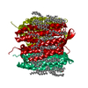
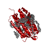
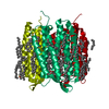
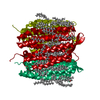
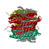
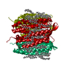
| Title | SCO6650, a 6-pyruvoyltetrahydropterin synthase homolog from Streptomyces coelicolor | ||||||
 Components Components | Uncharacterized protein SCO6650 | ||||||
 Keywords Keywords | UNKNOWN FUNCTION / T-fold | ||||||
| Function / homology |  Function and homology information Function and homology information 6-carboxytetrahydropterin synthase / 6-carboxy-5,6,7,8-tetrahydropterin synthase activity / metal ion binding 6-carboxytetrahydropterin synthase / 6-carboxy-5,6,7,8-tetrahydropterin synthase activity / metal ion bindingSimilarity search - Function | ||||||
| Biological species |  Streptomyces coelicolor (bacteria) Streptomyces coelicolor (bacteria) | ||||||
| Method | X-RAY DIFFRACTION / SYNCHROTRON / MAD / Resolution: 1.45 Å | ||||||
 Authors Authors | Spoonamore, J.E. / Roberts, S.A. / Heroux, A. / Bandarian, V. | ||||||
 Citation Citation | Journal: Acta Crystallogr.,Sect.F / Year: 2008 Title: Structure of a 6-pyruvoyltetrahydropterin synthase homolog from Streptomyces coelicolor. Authors: Spoonamore, J.E. / Roberts, S.A. / Heroux, A. / Bandarian, V. | ||||||
| History |
|
- Structure visualization
| Structure viewer | Molecule: MolmilJmol/JSmol |
|---|
- Downloads & links
-Download
| PDBx/mmCIF format | 3d7j.cif.gz | 176 KB | Display | PDBx/mmCIF format |
|---|---|---|---|---|
| PDB format | pdb3d7j.ent.gz | 148.7 KB | Display | PDB format |
| PDBx/mmJSON format | 3d7j.json.gz | Tree view | PDBx/mmJSON format | |
| Others |  Other downloads Other downloads |
-Validation report
| Arichive directory | https://data.pdbj.org/pub/pdb/validation_reports/d7/3d7jftp://data.pdbj.org/pub/pdb/validation_reports/d7/3d7j | HTTPS FTP |
|---|
-Related structure data
| Similar structure data |
|---|


-Links
 PDBj
PDBj
- Assembly
| Deposited unit | 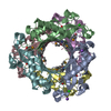
| ||||||||
|---|---|---|---|---|---|---|---|---|---|
| 1 |
| ||||||||
| Unit cell |
|
-Components
| #1: Protein | Mass: 16684.355 Da / Num. of mol.: 6 Source method: isolated from a genetically manipulated source Source: (gene. exp.) Streptomyces coelicolor (bacteria) / Strain: A(3)2 / Gene: SCO6650 / Plasmid: PET28 / Production host: Escherichia coli (E. coli) / Strain (production host): BL21(DE3) / References: UniProt: O86696#2: Chemical |   Mass: 22.990 Da / Num. of mol.: 2 / Source method: obtained synthetically / Formula: Na Mass: 22.990 Da / Num. of mol.: 2 / Source method: obtained synthetically / Formula: Na#3: Chemical | Chloride  Mass: 35.453 Da / Num. of mol.: 3 / Source method: obtained synthetically / Formula: Cl Mass: 35.453 Da / Num. of mol.: 3 / Source method: obtained synthetically / Formula: Cl#4: Water | ChemComp-HOH / | Water Mass: 18.015 Da / Num. of mol.: 677 / Source method: isolated from a natural source / Formula: H2O Mass: 18.015 Da / Num. of mol.: 677 / Source method: isolated from a natural source / Formula: H2O |
|---|
-Experimental details
-Experiment
| Experiment | Method: X-RAY DIFFRACTION / Number of used crystals: 2 |
|---|
- Sample preparation
| Crystal | Density Matthews: 2.06 Å3/Da / Density % sol: 40.19 % |
|---|---|
| Crystal grow | Temperature: 295 K / Method: vapor diffusion, hanging drop / pH: 8 Details: 2 M NaCl, 10% PEG6000, pH 8.0, VAPOR DIFFUSION, HANGING DROP, temperature 295K |
-Data collection
| Diffraction |
| ||||||||||||||||||
|---|---|---|---|---|---|---|---|---|---|---|---|---|---|---|---|---|---|---|---|
| Diffraction source |
| ||||||||||||||||||
| Detector |
| ||||||||||||||||||
| Radiation |
| ||||||||||||||||||
| Radiation wavelength |
| ||||||||||||||||||
| Reflection | Resolution: 1.45→41.34 Å / Num. all: 141171 / Num. obs: 140447 / % possible obs: 96 % / Observed criterion σ(F): 0 / Observed criterion σ(I): 0 / Redundancy: 6.5 % / Biso Wilson estimate: 19.3 Å2 / Rmerge(I) obs: 0.12 / Net I/σ(I): 44 | ||||||||||||||||||
| Reflection shell | Resolution: 1.45→1.48 Å / Redundancy: 2 % / Rmerge(I) obs: 0.49 / Mean I/σ(I) obs: 2.2 / Num. unique all: 7012 / % possible all: 72 |

- Processing
| Software |
| ||||||||||||||||||||||||||||||||||||||||||||||||||||||||||||||||||||||||||||||||||||||||||||||||||||||||||||||||||||||||||||||||||||||||||||||||||||||||||||||||||||||||||
|---|---|---|---|---|---|---|---|---|---|---|---|---|---|---|---|---|---|---|---|---|---|---|---|---|---|---|---|---|---|---|---|---|---|---|---|---|---|---|---|---|---|---|---|---|---|---|---|---|---|---|---|---|---|---|---|---|---|---|---|---|---|---|---|---|---|---|---|---|---|---|---|---|---|---|---|---|---|---|---|---|---|---|---|---|---|---|---|---|---|---|---|---|---|---|---|---|---|---|---|---|---|---|---|---|---|---|---|---|---|---|---|---|---|---|---|---|---|---|---|---|---|---|---|---|---|---|---|---|---|---|---|---|---|---|---|---|---|---|---|---|---|---|---|---|---|---|---|---|---|---|---|---|---|---|---|---|---|---|---|---|---|---|---|---|---|---|---|---|---|---|---|
| Refinement | Method to determine structure: MAD / Resolution: 1.45→41.34 Å / Cor.coef. Fo:Fc: 0.964 / Cor.coef. Fo:Fc free: 0.959 / SU B: 1.313 / SU ML: 0.052 / Cross valid method: THROUGHOUT / σ(F): 0 / ESU R: 0.08 / ESU R Free: 0.077 / Stereochemistry target values: MAXIMUM LIKELIHOOD / Details: HYDROGEN ATOMS HAVE BEEN ADDED IN RIDING POSITIONS
| ||||||||||||||||||||||||||||||||||||||||||||||||||||||||||||||||||||||||||||||||||||||||||||||||||||||||||||||||||||||||||||||||||||||||||||||||||||||||||||||||||||||||||
| Solvent computation | Ion probe radii: 0.8 Å / Shrinkage radii: 0.8 Å / VDW probe radii: 1.4 Å / Solvent model: MASK | ||||||||||||||||||||||||||||||||||||||||||||||||||||||||||||||||||||||||||||||||||||||||||||||||||||||||||||||||||||||||||||||||||||||||||||||||||||||||||||||||||||||||||
| Displacement parameters | Biso mean: 20.396 Å2
| ||||||||||||||||||||||||||||||||||||||||||||||||||||||||||||||||||||||||||||||||||||||||||||||||||||||||||||||||||||||||||||||||||||||||||||||||||||||||||||||||||||||||||
| Refinement step | Cycle: LAST / Resolution: 1.45→41.34 Å
| ||||||||||||||||||||||||||||||||||||||||||||||||||||||||||||||||||||||||||||||||||||||||||||||||||||||||||||||||||||||||||||||||||||||||||||||||||||||||||||||||||||||||||
| Refine LS restraints |
| ||||||||||||||||||||||||||||||||||||||||||||||||||||||||||||||||||||||||||||||||||||||||||||||||||||||||||||||||||||||||||||||||||||||||||||||||||||||||||||||||||||||||||
| LS refinement shell | Resolution: 1.45→1.485 Å / Total num. of bins used: 20
|
