 Movie
Movie Controller


+ Open data

- Basic information
| Entry | Database: PDB / ID: 6g70 | ||||||
|---|---|---|---|---|---|---|---|
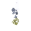
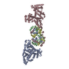
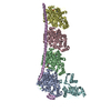
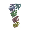
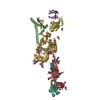
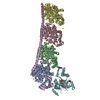
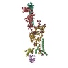
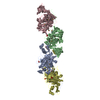
| Title | Structure of murine Prpf39 | ||||||
 Components Components | Pre-mRNA-processing factor 39 | ||||||
 Keywords Keywords |  SPLICING / U1snRNP / HAT repeat protein SPLICING / U1snRNP / HAT repeat protein | ||||||
| Function / homology |  Function and homology information Function and homology informationcommitment complex / U1 snRNP / U2-type prespliceosome / mRNA 5'-splice site recognition / RNA processingSimilarity search - Function | ||||||
| Biological species |  Mus musculus (house mouse) Mus musculus (house mouse) | ||||||
| Method | X-RAY DIFFRACTION / SYNCHROTRON / MOLECULAR REPLACEMENT / Resolution: 3.3 Å | ||||||
 Authors Authors | De Bortoli, F.D. / Loll, B. / Wahl, M. / Heyd, F. | ||||||
 Citation Citation | Journal: Nucleic Acids Res. / Year: 2019 Title: Increased versatility despite reduced molecular complexity: evolution, structure and function of metazoan splicing factor PRPF39. Authors: De Bortoli, F. / Neumann, A. / Kotte, A. / Timmermann, B. / Schuler, T. / Wahl, M.C. / Loll, B. / Heyd, F. | ||||||
| History |
|
- Structure visualization
| Structure viewer | Molecule: MolmilJmol/JSmol |
|---|
- Downloads & links
-Download
| PDBx/mmCIF format | 6g70.cif.gz | 441.8 KB | Display | PDBx/mmCIF format |
|---|---|---|---|---|
| PDB format | pdb6g70.ent.gz | 382.1 KB | Display | PDB format |
| PDBx/mmJSON format | 6g70.json.gz | Tree view | PDBx/mmJSON format | |
| Others |  Other downloads Other downloads |
-Validation report
| Arichive directory | https://data.pdbj.org/pub/pdb/validation_reports/g7/6g70ftp://data.pdbj.org/pub/pdb/validation_reports/g7/6g70 | HTTPS FTP |
|---|
-Related structure data
| Similar structure data | |
|---|---|
| Experimental dataset #1 | Data reference: 10.18430/m36g70 / Data set type: diffraction image data |
-Links
 PDBj
PDBj
- Assembly
| Deposited unit | 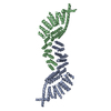
| ||||||||
|---|---|---|---|---|---|---|---|---|---|
| 1 |
| ||||||||
| Unit cell |
|
-Components
| #1: Protein | Mass: 78437.664 Da / Num. of mol.: 2 Source method: isolated from a genetically manipulated source Source: (gene. exp.) Mus musculus (house mouse) / Gene: Prpf39 / Plasmid: pGEX 6P1 / Production host:  Escherichia coli BL21(DE3) (bacteria) / Variant (production host): pLys / References: UniProt: E9QJV4, UniProt: Q8K2Z2*PLUS Escherichia coli BL21(DE3) (bacteria) / Variant (production host): pLys / References: UniProt: E9QJV4, UniProt: Q8K2Z2*PLUS |
|---|
-Experimental details
-Experiment
| Experiment | Method: X-RAY DIFFRACTION / Number of used crystals: 1 |
|---|
- Sample preparation
| Crystal | Density Matthews: 4.21 Å3/Da / Density % sol: 70.75 % |
|---|---|
| Crystal grow | Temperature: 277.15 K / Method: vapor diffusion, sitting drop / pH: 6.5 Details: 0.1 M bis-tris-propane pH 6.5, 1.8 M sodium acetate |
-Data collection
| Diffraction | Mean temperature: 100 K |
|---|---|
| Diffraction source | Source: SYNCHROTRON / Site: BESSY  / Beamline: 14.1 / Wavelength: 0.91841 Å / Beamline: 14.1 / Wavelength: 0.91841 Å |
| Detector | Type: DECTRIS PILATUS 6M / Detector: PIXEL / Date: Apr 19, 2016 |
| Radiation | Monochromator: SI111-DCM / Protocol: SINGLE WAVELENGTH / Monochromatic (M) / Laue (L): M / Scattering type: x-ray |
| Radiation wavelength | Wavelength: 0.91841 Å / Relative weight: 1 |
| Reflection | Resolution: 3.3→50 Å / Num. obs: 39170 / % possible obs: 98.5 % / Redundancy: 4.7 % / Biso Wilson estimate: 102 Å2 / CC1/2: 0.998 / Rrim(I) all: 0.149 / Net I/σ(I): 9.4 |
| Reflection shell | Resolution: 3.3→3.45 Å / Redundancy: 4.8 % / Mean I/σ(I) obs: 0.9 / Num. unique obs: 4809 / CC1/2: 0.637 / Rrim(I) all: 2.6 / % possible all: 97.9 |
- Processing
| Software |
| |||||||||||||||||||||||||||||||||||||||||||||||||||||||||||||||||||||||||||||||||||||||||||||||||||||||||||||||||||||||||||||||||||||||||||||||||||||||||||||||||||||||||||||||||||||||||||||||||||||||||||||||||||||||||||||||||
|---|---|---|---|---|---|---|---|---|---|---|---|---|---|---|---|---|---|---|---|---|---|---|---|---|---|---|---|---|---|---|---|---|---|---|---|---|---|---|---|---|---|---|---|---|---|---|---|---|---|---|---|---|---|---|---|---|---|---|---|---|---|---|---|---|---|---|---|---|---|---|---|---|---|---|---|---|---|---|---|---|---|---|---|---|---|---|---|---|---|---|---|---|---|---|---|---|---|---|---|---|---|---|---|---|---|---|---|---|---|---|---|---|---|---|---|---|---|---|---|---|---|---|---|---|---|---|---|---|---|---|---|---|---|---|---|---|---|---|---|---|---|---|---|---|---|---|---|---|---|---|---|---|---|---|---|---|---|---|---|---|---|---|---|---|---|---|---|---|---|---|---|---|---|---|---|---|---|---|---|---|---|---|---|---|---|---|---|---|---|---|---|---|---|---|---|---|---|---|---|---|---|---|---|---|---|---|---|---|---|---|---|---|---|---|---|---|---|---|---|---|---|---|---|---|---|---|
| Refinement | Method to determine structure: MOLECULAR REPLACEMENT / Resolution: 3.3→47.848 Å / SU ML: 0.61 / Cross valid method: FREE R-VALUE / σ(F): 1.33 / Phase error: 38.52
| |||||||||||||||||||||||||||||||||||||||||||||||||||||||||||||||||||||||||||||||||||||||||||||||||||||||||||||||||||||||||||||||||||||||||||||||||||||||||||||||||||||||||||||||||||||||||||||||||||||||||||||||||||||||||||||||||
| Solvent computation | Shrinkage radii: 0.9 Å / VDW probe radii: 1.11 Å | |||||||||||||||||||||||||||||||||||||||||||||||||||||||||||||||||||||||||||||||||||||||||||||||||||||||||||||||||||||||||||||||||||||||||||||||||||||||||||||||||||||||||||||||||||||||||||||||||||||||||||||||||||||||||||||||||
| Refinement step | Cycle: LAST / Resolution: 3.3→47.848 Å
| |||||||||||||||||||||||||||||||||||||||||||||||||||||||||||||||||||||||||||||||||||||||||||||||||||||||||||||||||||||||||||||||||||||||||||||||||||||||||||||||||||||||||||||||||||||||||||||||||||||||||||||||||||||||||||||||||
| Refine LS restraints |
| |||||||||||||||||||||||||||||||||||||||||||||||||||||||||||||||||||||||||||||||||||||||||||||||||||||||||||||||||||||||||||||||||||||||||||||||||||||||||||||||||||||||||||||||||||||||||||||||||||||||||||||||||||||||||||||||||
| LS refinement shell |
| |||||||||||||||||||||||||||||||||||||||||||||||||||||||||||||||||||||||||||||||||||||||||||||||||||||||||||||||||||||||||||||||||||||||||||||||||||||||||||||||||||||||||||||||||||||||||||||||||||||||||||||||||||||||||||||||||
| Refinement TLS params. | Method: refined / Refine-ID: X-RAY DIFFRACTION
| |||||||||||||||||||||||||||||||||||||||||||||||||||||||||||||||||||||||||||||||||||||||||||||||||||||||||||||||||||||||||||||||||||||||||||||||||||||||||||||||||||||||||||||||||||||||||||||||||||||||||||||||||||||||||||||||||
| Refinement TLS group |
|
