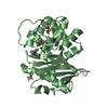
 Movie
Movie Controller


+ Open data

- Basic information
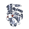
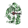
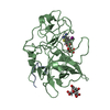
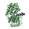
| Entry | Database: PDB / ID: 6g0j | ||||||
|---|---|---|---|---|---|---|---|
| Title | Inactive Fe-PP1 | ||||||
 Components Components | Serine/threonine-protein phosphatase PP1-alpha catalytic subunit | ||||||
 Keywords Keywords |  HYDROLASE / Phosphatase / Iron / oxidative stress / cell cycle HYDROLASE / Phosphatase / Iron / oxidative stress / cell cycle | ||||||
| Function / homology |  Function and homology information Function and homology informationregulation of glycogen catabolic process / PTW/PP1 phosphatase complex / glycogen granule / regulation of glycogen biosynthetic process / protein phosphatase 1 binding / cadherin binding involved in cell-cell adhesion / positive regulation of extrinsic apoptotic signaling pathway in absence of ligand / regulation of canonical Wnt signaling pathway / regulation of translational initiation / myosin phosphatase activity ...regulation of glycogen catabolic process / PTW/PP1 phosphatase complex / glycogen granule / regulation of glycogen biosynthetic process / protein phosphatase 1 binding / cadherin binding involved in cell-cell adhesion / positive regulation of extrinsic apoptotic signaling pathway in absence of ligand / regulation of canonical Wnt signaling pathway / regulation of translational initiation / myosin phosphatase activity / protein serine/threonine phosphatase activity / branching morphogenesis of an epithelial tube / glycogen metabolic process / protein-serine/threonine phosphatase / entrainment of circadian clock by photoperiod / Triglyceride catabolism / phosphatase activity / phosphoprotein phosphatase activity / DARPP-32 events / ribonucleoprotein complex binding / dephosphorylation / protein dephosphorylation / Downregulation of TGF-beta receptor signaling / response to lead ion / adherens junction / lung development / circadian regulation of gene expression / regulation of circadian rhythm / Circadian Clock / presynapse / perikaryon / dendritic spine / cell cycle / cell division / glutamatergic synapse / nucleolus / extracellular exosome / nucleoplasm / metal ion binding / nucleus / plasma membrane / cytosol / cytoplasmSimilarity search - Function | ||||||
| Biological species |  Homo sapiens (human) Homo sapiens (human) | ||||||
| Method | X-RAY DIFFRACTION / SYNCHROTRON / MOLECULAR REPLACEMENT / Resolution: 2.1 Å | ||||||
 Authors Authors | Salvi, F. / Barabas, O. / Koehn, M. | ||||||
| Funding support |  Germany, 1items Germany, 1items
| ||||||
 Citation Citation | Journal: FEBS Lett. / Year: 2018 Title: Effects of stably incorporated iron on protein phosphatase-1 structure and activity. Authors: Salvi, F. / Trebacz, M. / Kokot, T. / Hoermann, B. / Rios, P. / Barabas, O. / Koehn, M. | ||||||
| History |
|
- Structure visualization
| Structure viewer | Molecule: MolmilJmol/JSmol |
|---|
- Downloads & links
-Download
| PDBx/mmCIF format | 6g0j.cif.gz | 77.2 KB | Display | PDBx/mmCIF format |
|---|---|---|---|---|
| PDB format | pdb6g0j.ent.gz | 54.8 KB | Display | PDB format |
| PDBx/mmJSON format | 6g0j.json.gz | Tree view | PDBx/mmJSON format | |
| Others |  Other downloads Other downloads |
-Validation report
| Arichive directory | https://data.pdbj.org/pub/pdb/validation_reports/g0/6g0jftp://data.pdbj.org/pub/pdb/validation_reports/g0/6g0j | HTTPS FTP |
|---|
-Related structure data
| Related structure data | 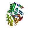 6g0iC  4movS S: Starting model for refinement C: citing same article ( |
|---|---|
| Similar structure data |


-Links
 PDBj
PDBj


- Assembly
| Deposited unit | 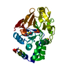
| ||||||||
|---|---|---|---|---|---|---|---|---|---|
| 1 |
| ||||||||
| Unit cell |
|
-Components
| #1: Protein | Mass: 37647.102 Da / Num. of mol.: 1 Source method: isolated from a genetically manipulated source Details: Two cysteine residues are modeled as oxidized / Source: (gene. exp.) Homo sapiens (human) / Gene: PPP1CA, PPP1A / Production host:  Escherichia coli (E. coli) Escherichia coli (E. coli)References: UniProt: P62136, protein-serine/threonine phosphatase | ||||||
|---|---|---|---|---|---|---|---|
| #2: Chemical | Iron  Mass: 55.845 Da / Num. of mol.: 2 / Source method: obtained synthetically / Formula: Fe / Feature type: SUBJECT OF INVESTIGATION Mass: 55.845 Da / Num. of mol.: 2 / Source method: obtained synthetically / Formula: Fe / Feature type: SUBJECT OF INVESTIGATION#3: Chemical |   Mass: 54.938 Da / Num. of mol.: 2 / Source method: obtained synthetically / Formula: Mn / Feature type: SUBJECT OF INVESTIGATION Mass: 54.938 Da / Num. of mol.: 2 / Source method: obtained synthetically / Formula: Mn / Feature type: SUBJECT OF INVESTIGATION#4: Chemical | ChemComp-PO4 / | Phosphate  Mass: 94.971 Da / Num. of mol.: 1 / Source method: obtained synthetically / Formula: PO4 / Feature type: SUBJECT OF INVESTIGATION Mass: 94.971 Da / Num. of mol.: 1 / Source method: obtained synthetically / Formula: PO4 / Feature type: SUBJECT OF INVESTIGATION#5: Water | ChemComp-HOH / | Water Mass: 18.015 Da / Num. of mol.: 114 / Source method: isolated from a natural source / Formula: H2O Mass: 18.015 Da / Num. of mol.: 114 / Source method: isolated from a natural source / Formula: H2O |
-Experimental details
-Experiment
| Experiment | Method: X-RAY DIFFRACTION / Number of used crystals: 1 |
|---|
- Sample preparation
| Crystal | Density Matthews: 2.57 Å3/Da / Density % sol: 52.15 % |
|---|---|
| Crystal grow | Temperature: 291 K / Method: vapor diffusion, sitting drop / pH: 8 Details: 28% w/v PEG 3350, 0.1 M TRIS-Cl,pH 8.0 RT, 1 M Lithium Chloride. |
-Data collection
| Diffraction | Mean temperature: 100 K |
|---|---|
| Diffraction source | Source: SYNCHROTRON / Site: ESRF  / Beamline: MASSIF-3 / Wavelength: 0.9677 Å / Beamline: MASSIF-3 / Wavelength: 0.9677 Å |
| Detector | Type: DECTRIS EIGER X 4M / Detector: PIXEL / Date: Nov 30, 2016 |
| Radiation | Protocol: SINGLE WAVELENGTH / Monochromatic (M) / Laue (L): M / Scattering type: x-ray |
| Radiation wavelength | Wavelength: 0.9677 Å / Relative weight: 1 |
| Reflection | Resolution: 2.1→46.81 Å / Num. obs: 19896 / % possible obs: 97.3 % / Redundancy: 4.1 % / CC1/2: 0.998 / Rmerge(I) obs: 0.068 / Net I/σ(I): 12.9 |
| Reflection shell | Resolution: 2.1→2.16 Å / Redundancy: 4.2 % / Rmerge(I) obs: 0.545 / Mean I/σ(I) obs: 2.3 / Num. unique obs: 1662 / CC1/2: 0.806 / % possible all: 98.9 |
- Processing
| Software |
| ||||||||||||||||||||||||||||||||||||||||||||||||||||||||||||||||||||||||||||||||||||||||||||||||||||||||||||||||||||||||||||||||||||||||||||||||||||||||||||||||||||||||||||||||||||||
|---|---|---|---|---|---|---|---|---|---|---|---|---|---|---|---|---|---|---|---|---|---|---|---|---|---|---|---|---|---|---|---|---|---|---|---|---|---|---|---|---|---|---|---|---|---|---|---|---|---|---|---|---|---|---|---|---|---|---|---|---|---|---|---|---|---|---|---|---|---|---|---|---|---|---|---|---|---|---|---|---|---|---|---|---|---|---|---|---|---|---|---|---|---|---|---|---|---|---|---|---|---|---|---|---|---|---|---|---|---|---|---|---|---|---|---|---|---|---|---|---|---|---|---|---|---|---|---|---|---|---|---|---|---|---|---|---|---|---|---|---|---|---|---|---|---|---|---|---|---|---|---|---|---|---|---|---|---|---|---|---|---|---|---|---|---|---|---|---|---|---|---|---|---|---|---|---|---|---|---|---|---|---|---|
| Refinement | Method to determine structure: MOLECULAR REPLACEMENT Starting model: 4MOV Resolution: 2.1→46.81 Å / Cor.coef. Fo:Fc: 0.952 / Cor.coef. Fo:Fc free: 0.934 / SU B: 5.442 / SU ML: 0.135 / Cross valid method: THROUGHOUT / ESU R: 0.219 / ESU R Free: 0.181 / Details: HYDROGENS HAVE BEEN ADDED IN THE RIDING POSITIONS
| ||||||||||||||||||||||||||||||||||||||||||||||||||||||||||||||||||||||||||||||||||||||||||||||||||||||||||||||||||||||||||||||||||||||||||||||||||||||||||||||||||||||||||||||||||||||
| Solvent computation | Ion probe radii: 0.8 Å / Shrinkage radii: 0.8 Å / VDW probe radii: 1.2 Å | ||||||||||||||||||||||||||||||||||||||||||||||||||||||||||||||||||||||||||||||||||||||||||||||||||||||||||||||||||||||||||||||||||||||||||||||||||||||||||||||||||||||||||||||||||||||
| Displacement parameters | Biso mean: 36.462 Å2
| ||||||||||||||||||||||||||||||||||||||||||||||||||||||||||||||||||||||||||||||||||||||||||||||||||||||||||||||||||||||||||||||||||||||||||||||||||||||||||||||||||||||||||||||||||||||
| Refinement step | Cycle: 1 / Resolution: 2.1→46.81 Å
| ||||||||||||||||||||||||||||||||||||||||||||||||||||||||||||||||||||||||||||||||||||||||||||||||||||||||||||||||||||||||||||||||||||||||||||||||||||||||||||||||||||||||||||||||||||||
| Refine LS restraints |
|