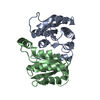
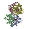
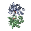
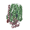
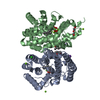
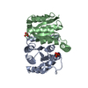
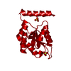
[English] 日本語
Yorodumi
- PDB-6fxw: Structure of Leishmania infantum type B ribose 5-phosphate isomerase -

+
Open data


ID or keywords:

Loading...

-
Basic information

Entry
Database: PDB / ID: 6fxw
TitleStructure of Leishmania infantum type B ribose 5-phosphate isomerase
ComponentsPutative ribose 5-phosphate isomerase
KeywordsISOMERASE / PENTOSE PHOSPHATE PATHWAY TYPE B RIBOSE 5-PHOSPHATE ISOMERASE R5P
Function / homology
Function and homology information


ribose-5-phosphate isomerase / ribose-5-phosphate isomerase activity / carbohydrate metabolic process
Similarity search - Function
Ribose 5-phosphate isomerase B / Sugar-phosphate isomerase, RpiB/LacA/LacB family / Sugar-phosphate isomerase, RpiB/LacA/LacB superfamily / Ribose/Galactose Isomerase / Ribose 5-phosphate Isomerase B; Chain: A, / Sugar-phosphate isomerase, RpiB/LacA/LacB / 3-Layer(aba) Sandwich / Alpha Beta
Similarity search - Domain/homology
Putative ribose 5-phosphate isomerase
Similarity search - Component
Biological speciesLeishmania infantum (eukaryote)
MethodX-RAY DIFFRACTION / SYNCHROTRON / MOLECULAR REPLACEMENT / Resolution: 1.57 Å
AuthorsRonin, C. / Ciesielski, F. / Ciapetti, P.
Funding support France, 1items
OrganizationGrant numberCountry
European Communitys Seventh Framework Programme602773 France
CitationJournal: Antimicrob.Agents Chemother. / Year: 2021
Title: Toward Chemical Validation of Leishmania infantum Ribose 5-Phosphate Isomerase as a Drug Target.
Authors: Dickie, E.A. / Ronin, C. / Sa, M. / Ciesielski, F. / Trouche, N. / Tavares, J. / Santarem, N. / Major, L.L. / Pemberton, I.K. / MacDougall, J. / Smith, T.K. / Cordeiro-da-Silva, A. / Ciapetti, P.
History
DepositionMar 9, 2018Deposition site: PDBE / Processing site: PDBE
Revision 1.0May 22, 2019Provider: repository / Type: Initial release
Revision 1.1Jun 12, 2019Group: Data collection / Database references / Structure summary
Category: audit_author / citation_author ...audit_author / citation_author / database_PDB_rev / database_PDB_rev_record
Item: _audit_author.name / _citation_author.name
Revision 1.2Jan 17, 2024Group: Author supporting evidence / Data collection ...Author supporting evidence / Data collection / Database references / Refinement description
Category: chem_comp_atom / chem_comp_bond ...chem_comp_atom / chem_comp_bond / database_2 / pdbx_audit_support / pdbx_initial_refinement_model
Item: _database_2.pdbx_DOI / _database_2.pdbx_database_accession / _pdbx_audit_support.funding_organization
Revision 1.3Jun 12, 2024Group: Database references / Category: citation / citation_author
Item: _citation.country / _citation.journal_abbrev ..._citation.country / _citation.journal_abbrev / _citation.journal_id_ASTM / _citation.journal_id_CSD / _citation.journal_id_ISSN / _citation.journal_volume / _citation.page_first / _citation.page_last / _citation.pdbx_database_id_DOI / _citation.pdbx_database_id_PubMed / _citation.title / _citation.year

-
Structure visualization

Structure viewerMolecule:
MolmilJmol/JSmol

Downloads & links

-
Assembly

Deposited unit
A: Putative ribose 5-phosphate isomerase
hetero molecules


Theoretical massNumber of molelcules
Total (without water)19,1663
Polymers18,9741
Non-polymers1922
Water1,65792
1
A: Putative ribose 5-phosphate isomerase
hetero molecules

A: Putative ribose 5-phosphate isomerase
hetero molecules

A: Putative ribose 5-phosphate isomerase
hetero molecules

A: Putative ribose 5-phosphate isomerase
hetero molecules


Theoretical massNumber of molelcules
Total (without water)76,66312
Polymers75,8944
Non-polymers7698
Water724
TypeNameSymmetry operationNumber
identity operation1_555x,y,z1
crystal symmetry operation8_554x,-y+1/2,-z-1/21
crystal symmetry operation11_454-x-1/2,y,-z-1/21
crystal symmetry operation14_455-x-1/2,-y+1/2,z1
Buried area10820 Å2
ΔGint-148 kcal/mol
Surface area21810 Å2
MethodPISA
Unit cell
Length a, b, c (Å)80.920, 83.650, 89.490
Angle α, β, γ (deg.)90.00, 90.00, 90.00
Int Tables number22
Space group name H-MF222
Components on special symmetry positions
IDModelComponents
11A-305-

HOH

21A-390-

HOH

31A-392-

HOH

-
Components

#1: Protein Putative ribose 5-phosphate isomerase


Mass: 18973.559 Da / Num. of mol.: 1
Source method: isolated from a genetically manipulated source
Source: (gene. exp.) Leishmania infantum (eukaryote) / Gene: LINJ_28_2100 / Production host: Escherichia coli (E. coli) / References: UniProt: A4I3S4, ribose-5-phosphate isomerase
#2: Chemical ChemComp-SO4 / SULFATE ION / Sulfate


Mass: 96.063 Da / Num. of mol.: 2 / Source method: obtained synthetically / Formula: SO4
#3: Water ChemComp-HOH / water / Water


Mass: 18.015 Da / Num. of mol.: 92 / Source method: isolated from a natural source / Formula: H2O

-
Experimental details

-
Experiment

ExperimentMethod: X-RAY DIFFRACTION / Number of used crystals: 1

-
Sample preparation

CrystalDensity Matthews: 2 Å3/Da / Density % sol: 38.36 %
Crystal growTemperature: 277 K / Method: vapor diffusion, sitting drop / Details: 0.1M Hepes pH7; 2M NH4SO4

-
Data collection

DiffractionMean temperature: 100 K
Diffraction sourceSource: SYNCHROTRON / Site: SOLEIL / Beamline: PROXIMA 1 / Wavelength: 0.912 Å
DetectorType: DECTRIS PILATUS 6M / Detector: PIXEL / Date: Jul 21, 2015
RadiationProtocol: SINGLE WAVELENGTH / Monochromatic (M) / Laue (L): M / Scattering type: x-ray
Radiation wavelengthWavelength: 0.912 Å / Relative weight: 1
ReflectionResolution: 1.57→19.75 Å / Num. obs: 21306 / % possible obs: 99.6 % / Redundancy: 6.5 % / Rsym value: 0.0527 / Net I/σ(I): 23.2
Reflection shellResolution: 1.57→1.66 Å / Mean I/σ(I) obs: 5.09 / Rsym value: 0.343 / % possible all: 97.8

-
Processing

Software
NameVersionClassification
REFMAC5.8.0189refinement
XDSdata reduction
XDSdata scaling
PHASERphasing
RefinementMethod to determine structure: MOLECULAR REPLACEMENT
Starting model: 3k7o
Resolution: 1.57→19.75 Å / Cor.coef. Fo:Fc: 0.973 / Cor.coef. Fo:Fc free: 0.961 / SU B: 1.207 / SU ML: 0.043 / Cross valid method: THROUGHOUT / ESU R: 0.067 / ESU R Free: 0.071 / Stereochemistry target values: MAXIMUM LIKELIHOOD / Details: HYDROGENS HAVE BEEN ADDED IN THE RIDING POSITIONS
RfactorNum. reflection% reflectionSelection details
Rfree0.17829 1066 5 %RANDOM
Rwork0.14439 ---
obs0.14611 20240 99.55 %-
Solvent computationIon probe radii: 0.8 Å / Shrinkage radii: 0.8 Å / VDW probe radii: 1.2 Å / Solvent model: MASK
Displacement parametersBiso mean: 17.348 Å2
Baniso -1Baniso -2Baniso -3
1-0.28 Å20 Å20 Å2
2--0.32 Å20 Å2
3----0.59 Å2
Refinement stepCycle: 1 / Resolution: 1.57→19.75 Å
ProteinNucleic acidLigandSolventTotal
Num. atoms1149 0 10 92 1251
Refine LS restraints
Refine-IDTypeDev idealDev ideal targetNumber
X-RAY DIFFRACTIONr_bond_refined_d0.0240.0191202
X-RAY DIFFRACTIONr_bond_other_d0.0020.021114
X-RAY DIFFRACTIONr_angle_refined_deg2.2171.9611635
X-RAY DIFFRACTIONr_angle_other_deg1.13932590
X-RAY DIFFRACTIONr_dihedral_angle_1_deg6.225164
X-RAY DIFFRACTIONr_dihedral_angle_2_deg35.65724.250
X-RAY DIFFRACTIONr_dihedral_angle_3_deg12.16215210
X-RAY DIFFRACTIONr_dihedral_angle_4_deg15.196158
X-RAY DIFFRACTIONr_chiral_restr0.1370.2192
X-RAY DIFFRACTIONr_gen_planes_refined0.0110.021356
X-RAY DIFFRACTIONr_gen_planes_other0.0030.02228
X-RAY DIFFRACTIONr_nbd_refined
X-RAY DIFFRACTIONr_nbd_other
X-RAY DIFFRACTIONr_nbtor_refined
X-RAY DIFFRACTIONr_nbtor_other
X-RAY DIFFRACTIONr_xyhbond_nbd_refined
X-RAY DIFFRACTIONr_xyhbond_nbd_other
X-RAY DIFFRACTIONr_metal_ion_refined
X-RAY DIFFRACTIONr_metal_ion_other
X-RAY DIFFRACTIONr_symmetry_vdw_refined
X-RAY DIFFRACTIONr_symmetry_vdw_other
X-RAY DIFFRACTIONr_symmetry_hbond_refined
X-RAY DIFFRACTIONr_symmetry_hbond_other
X-RAY DIFFRACTIONr_symmetry_metal_ion_refined
X-RAY DIFFRACTIONr_symmetry_metal_ion_other
X-RAY DIFFRACTIONr_mcbond_it2.0461.526623
X-RAY DIFFRACTIONr_mcbond_other1.9591.518622
X-RAY DIFFRACTIONr_mcangle_it2.9472.277780
X-RAY DIFFRACTIONr_mcangle_other2.9582.285781
X-RAY DIFFRACTIONr_scbond_it3.211.878579
X-RAY DIFFRACTIONr_scbond_other3.2151.872572
X-RAY DIFFRACTIONr_scangle_it
X-RAY DIFFRACTIONr_scangle_other4.8312.663838
X-RAY DIFFRACTIONr_long_range_B_refined5.70119.1211330
X-RAY DIFFRACTIONr_long_range_B_other5.69118.9431319
X-RAY DIFFRACTIONr_rigid_bond_restr
X-RAY DIFFRACTIONr_sphericity_free
X-RAY DIFFRACTIONr_sphericity_bonded
LS refinement shellResolution: 1.569→1.609 Å / Total num. of bins used: 20
RfactorNum. reflection% reflection
Rfree0.32 74 -
Rwork0.269 1407 -
obs--94.94 %

+
About Yorodumi

-
News

-
Feb 9, 2022. New format data for meta-information of EMDB entries

New format data for meta-information of EMDB entries

  • Version 3 of the EMDB header file is now the official format.
  • The previous official version 1.9 will be removed from the archive.

Related info.:EMDB header

External links:wwPDB to switch to version 3 of the EMDB data model

-
Aug 12, 2020. Covid-19 info

Covid-19 info

URL: https://pdbj.org/emnavi/covid19.php

New page: Covid-19 featured information page in EM Navigator.

Related info.:Covid-19 info / Mar 5, 2020. Novel coronavirus structure data

+
Mar 5, 2020. Novel coronavirus structure data

Novel coronavirus structure data

Related info.:Yorodumi Speices / Aug 12, 2020. Covid-19 info

External links:COVID-19 featured content - PDBj / Molecule of the Month (242):Coronavirus Proteases

+
Jan 31, 2019. EMDB accession codes are about to change! (news from PDBe EMDB page)

EMDB accession codes are about to change! (news from PDBe EMDB page)

  • The allocation of 4 digits for EMDB accession codes will soon come to an end. Whilst these codes will remain in use, new EMDB accession codes will include an additional digit and will expand incrementally as the available range of codes is exhausted. The current 4-digit format prefixed with “EMD-” (i.e. EMD-XXXX) will advance to a 5-digit format (i.e. EMD-XXXXX), and so on. It is currently estimated that the 4-digit codes will be depleted around Spring 2019, at which point the 5-digit format will come into force.
  • The EM Navigator/Yorodumi systems omit the EMD- prefix.

Related info.:Q: What is EMD? / ID/Accession-code notation in Yorodumi/EM Navigator

External links:EMDB Accession Codes are Changing Soon! / Contact to PDBj

+
Jul 12, 2017. Major update of PDB

Major update of PDB

  • wwPDB released updated PDB data conforming to the new PDBx/mmCIF dictionary.
  • This is a major update changing the version number from 4 to 5, and with Remediation, in which all the entries are updated.
  • In this update, many items about electron microscopy experimental information are reorganized (e.g. em_software).
  • Now, EM Navigator and Yorodumi are based on the updated data.

External links:wwPDB Remediation / Enriched Model Files Conforming to OneDep Data Standards Now Available in the PDB FTP Archive

-
Yorodumi

Thousand views of thousand structures

  • Yorodumi is a browser for structure data from EMDB, PDB, SASBDB, etc.
  • This page is also the successor to EM Navigator detail page, and also detail information page/front-end page for Omokage search.
  • The word "yorodu" (or yorozu) is an old Japanese word meaning "ten thousand". "mi" (miru) is to see.

Related info.:EMDB / PDB / SASBDB / Comparison of 3 databanks / Yorodumi Search / Aug 31, 2016. New EM Navigator & Yorodumi / Yorodumi Papers / Jmol/JSmol / Function and homology information / Changes in new EM Navigator and Yorodumi

Read more