 Movie
Movie Controller

[English] 日本語
 Yorodumi
- PDB-3m1p: Structure of ribose 5-phosphate isomerase type B from Trypanosoma... -
+ Open data

- Basic information
| Entry | Database: PDB / ID: 3m1p | ||||||
|---|---|---|---|---|---|---|---|
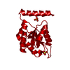
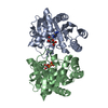
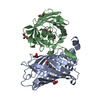
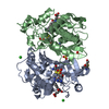
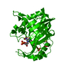
| Title | Structure of ribose 5-phosphate isomerase type B from Trypanosoma cruzi, soaked with allose-6-phosphate | ||||||
 Components Components | Ribose 5-phosphate isomerase | ||||||
 Keywords Keywords |  ISOMERASE / type B ribose 5-phosphate isomerase / allose-6-phosphate ISOMERASE / type B ribose 5-phosphate isomerase / allose-6-phosphate | ||||||
| Function / homology |  Function and homology informationisomerase activity / carbohydrate metabolic process / identical protein binding Function and homology informationisomerase activity / carbohydrate metabolic process / identical protein bindingSimilarity search - Function | ||||||
| Biological species |  Trypanosoma cruzi (eukaryote) Trypanosoma cruzi (eukaryote) | ||||||
| Method | X-RAY DIFFRACTION / SYNCHROTRON / MOLECULAR REPLACEMENT / Resolution: 2.2 Å | ||||||
 Authors Authors | Naworyta, A. / Mowbray, S.L. / Stern, A.L. | ||||||
 Citation Citation | Journal: Febs J. / Year: 2011 Title: Structures of type B ribose 5-phosphate isomerase from Trypanosoma cruzi shed light on the determinants of sugar specificity in the structural family. Authors: Stern, A.L. / Naworyta, A. / Cazzulo, J.J. / Mowbray, S.L. | ||||||
| History |
|
- Structure visualization
| Structure viewer | Molecule: MolmilJmol/JSmol |
|---|
- Downloads & links
-Download
| PDBx/mmCIF format | 3m1p.cif.gz | 72.2 KB | Display | PDBx/mmCIF format |
|---|---|---|---|---|
| PDB format | pdb3m1p.ent.gz | 55.4 KB | Display | PDB format |
| PDBx/mmJSON format | 3m1p.json.gz | Tree view | PDBx/mmJSON format | |
| Others |  Other downloads Other downloads |
-Validation report
| Arichive directory | https://data.pdbj.org/pub/pdb/validation_reports/m1/3m1pftp://data.pdbj.org/pub/pdb/validation_reports/m1/3m1p | HTTPS FTP |
|---|
-Related structure data


-Links
 PDBj
PDBj- Assembly
| Deposited unit | 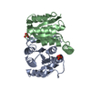
| ||||||||
|---|---|---|---|---|---|---|---|---|---|
| 1 | 
| ||||||||
| Unit cell |
|
-Components
| #1: Protein | Mass: 16756.166 Da / Num. of mol.: 2 / Mutation: C69A Source method: isolated from a genetically manipulated source Source: (gene. exp.) Trypanosoma cruzi (eukaryote) / Gene: Tc00.1047053509199.24 / Production host:  Escherichia coli (E. coli) / References: UniProt: Q4CQE2, ribose-5-phosphate isomerase Escherichia coli (E. coli) / References: UniProt: Q4CQE2, ribose-5-phosphate isomerase#2: Chemical | Phosphate  Mass: 94.971 Da / Num. of mol.: 2 / Source method: obtained synthetically / Formula: PO4 Mass: 94.971 Da / Num. of mol.: 2 / Source method: obtained synthetically / Formula: PO4#3: Water | ChemComp-HOH / | Water Mass: 18.015 Da / Num. of mol.: 136 / Source method: isolated from a natural source / Formula: H2O Mass: 18.015 Da / Num. of mol.: 136 / Source method: isolated from a natural source / Formula: H2O |
|---|
-Experimental details
-Experiment
| Experiment | Method: X-RAY DIFFRACTION / Number of used crystals: 1 |
|---|
- Sample preparation
| Crystal | Density Matthews: 2.99 Å3/Da / Density % sol: 58.83 % |
|---|---|
| Crystal grow | Temperature: 293 K / Method: vapor diffusion / pH: 8.5 Details: 20% PEG3350, 0.2M sodium acetate, 0.1 M BIS-TRIS propane, pH 8.5, VAPOR DIFFUSION, temperature 293K |
-Data collection
| Diffraction | Mean temperature: 100 K | ||||||||||||||||||||||||||||||||||||||||||||||||||||||||||||||||||||||||||||||||||||||||
|---|---|---|---|---|---|---|---|---|---|---|---|---|---|---|---|---|---|---|---|---|---|---|---|---|---|---|---|---|---|---|---|---|---|---|---|---|---|---|---|---|---|---|---|---|---|---|---|---|---|---|---|---|---|---|---|---|---|---|---|---|---|---|---|---|---|---|---|---|---|---|---|---|---|---|---|---|---|---|---|---|---|---|---|---|---|---|---|---|---|
| Diffraction source | Source: SYNCHROTRON / Site: ESRF  / Beamline: BM30A / Beamline: BM30A | ||||||||||||||||||||||||||||||||||||||||||||||||||||||||||||||||||||||||||||||||||||||||
| Detector | Type: ADSC QUANTUM 315r / Detector: CCD / Date: Feb 8, 2009 | ||||||||||||||||||||||||||||||||||||||||||||||||||||||||||||||||||||||||||||||||||||||||
| Radiation | Protocol: SINGLE WAVELENGTH / Monochromatic (M) / Laue (L): M / Scattering type: x-ray | ||||||||||||||||||||||||||||||||||||||||||||||||||||||||||||||||||||||||||||||||||||||||
| Radiation wavelength | Relative weight: 1 | ||||||||||||||||||||||||||||||||||||||||||||||||||||||||||||||||||||||||||||||||||||||||
| Reflection | Resolution: 2.2→65.71 Å / Num. all: 18940 / Num. obs: 18940 / % possible obs: 90.1 % / Redundancy: 6.5 % / Rmerge(I) obs: 0.105 / Rsym value: 0.105 / Net I/σ(I): 16.3 | ||||||||||||||||||||||||||||||||||||||||||||||||||||||||||||||||||||||||||||||||||||||||
| Reflection shell |
|
- Processing
| Software |
| ||||||||||||||||||||||||||||||||||||||||||||||||||||||||||||||||||||||||||||||||||||||||||
|---|---|---|---|---|---|---|---|---|---|---|---|---|---|---|---|---|---|---|---|---|---|---|---|---|---|---|---|---|---|---|---|---|---|---|---|---|---|---|---|---|---|---|---|---|---|---|---|---|---|---|---|---|---|---|---|---|---|---|---|---|---|---|---|---|---|---|---|---|---|---|---|---|---|---|---|---|---|---|---|---|---|---|---|---|---|---|---|---|---|---|---|
| Refinement | Method to determine structure: MOLECULAR REPLACEMENT / Resolution: 2.2→30 Å / Cor.coef. Fo:Fc: 0.939 / Cor.coef. Fo:Fc free: 0.912 / WRfactor Rfree: 0.214 / WRfactor Rwork: 0.175 / Occupancy max: 1 / Occupancy min: 0.5 / FOM work R set: 0.863 / SU B: 4.712 / SU ML: 0.122 / SU R Cruickshank DPI: 0.253 / SU Rfree: 0.205 / Cross valid method: THROUGHOUT / σ(F): 0 / ESU R: 0.253 / ESU R Free: 0.205 / Stereochemistry target values: MAXIMUM LIKELIHOOD / Details: HYDROGENS HAVE BEEN ADDED IN THE RIDING POSITIONS
| ||||||||||||||||||||||||||||||||||||||||||||||||||||||||||||||||||||||||||||||||||||||||||
| Solvent computation | Ion probe radii: 0.8 Å / Shrinkage radii: 0.8 Å / VDW probe radii: 1.2 Å / Solvent model: MASK | ||||||||||||||||||||||||||||||||||||||||||||||||||||||||||||||||||||||||||||||||||||||||||
| Displacement parameters | Biso max: 48.76 Å2 / Biso mean: 21.072 Å2 / Biso min: 6.58 Å2
| ||||||||||||||||||||||||||||||||||||||||||||||||||||||||||||||||||||||||||||||||||||||||||
| Refinement step | Cycle: LAST / Resolution: 2.2→30 Å
| ||||||||||||||||||||||||||||||||||||||||||||||||||||||||||||||||||||||||||||||||||||||||||
| Refine LS restraints |
| ||||||||||||||||||||||||||||||||||||||||||||||||||||||||||||||||||||||||||||||||||||||||||
| LS refinement shell | Resolution: 2.2→2.257 Å / Total num. of bins used: 20
|
