 Movie
Movie Controller

[English] 日本語
 Yorodumi
- PDB-6fxe: BBE31 from Lyme disease agent Borrelia (Borreliella) burgdorferi ... -
+ Open data

- Basic information
| Entry | Database: PDB / ID: 6fxe | ||||||
|---|---|---|---|---|---|---|---|
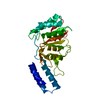
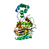
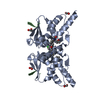
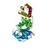
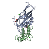
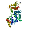
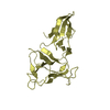
| Title | BBE31 from Lyme disease agent Borrelia (Borreliella) burgdorferi playing a vital role in successful colonization of the mammalian host | ||||||
 Components Components | Putative surface protein Cell membrane Cell membrane | ||||||
 Keywords Keywords | PROTEIN BINDING / Outer surface protein / borrelia outer membrane / pFam54 family / glutathione binding | ||||||
| Function / homology | Borrelia lipoprotein paralogus family 54/60 / Borrelia Bbcrasp-1 domain containing protein / GLUTATHIONE / Putative surface protein Function and homology information Function and homology information | ||||||
| Biological species |  Borrelia burgdorferi B31 (bacteria) Borrelia burgdorferi B31 (bacteria) | ||||||
| Method | X-RAY DIFFRACTION / SYNCHROTRON / SAD / Resolution: 2.4 Å | ||||||
 Authors Authors | Brangulis, K. / Akopjana, I. / Petrovskis, I. / Kazaks, A. / Tars, K. | ||||||
| Funding support |  Latvia, 1items Latvia, 1items
| ||||||
 Citation Citation | Journal: Biochim Biophys Acta Gen Subj / Year: 2019 Title: BBE31 from the Lyme disease agent Borrelia burgdorferi, known to play an important role in successful colonization of the mammalian host, shows the ability to bind glutathione. Authors: Brangulis, K. / Akopjana, I. / Petrovskis, I. / Kazaks, A. / Zelencova, D. / Jekabsons, A. / Jaudzems, K. / Tars, K. | ||||||
| History |
|
- Structure visualization
| Structure viewer | Molecule: MolmilJmol/JSmol |
|---|
- Downloads & links
-Download
| PDBx/mmCIF format | 6fxe.cif.gz | 89.8 KB | Display | PDBx/mmCIF format |
|---|---|---|---|---|
| PDB format | pdb6fxe.ent.gz | 73.1 KB | Display | PDB format |
| PDBx/mmJSON format | 6fxe.json.gz | Tree view | PDBx/mmJSON format | |
| Others |  Other downloads Other downloads |
-Validation report
| Arichive directory | https://data.pdbj.org/pub/pdb/validation_reports/fx/6fxeftp://data.pdbj.org/pub/pdb/validation_reports/fx/6fxe | HTTPS FTP |
|---|
-Related structure data


-Links
 PDBj
PDBj

- Assembly
| Deposited unit | 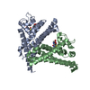
| ||||||||
|---|---|---|---|---|---|---|---|---|---|
| 1 | 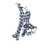
| ||||||||
| 2 | 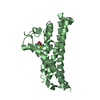
| ||||||||
| Unit cell |
|
-Components
| #1: Protein | Cell membrane Mass: 24255.869 Da / Num. of mol.: 2 Source method: isolated from a genetically manipulated source Details: First four amino acids (GAMG) are remnants from the expression tag. Source: (gene. exp.) Borrelia burgdorferi B31 (bacteria) / Gene: BB_E31 / Production host: Escherichia coli BL21 (bacteria) / References: UniProt: O50725#2: Chemical | Glutathione  Mass: 307.323 Da / Num. of mol.: 2 / Source method: obtained synthetically / Formula: C10H17N3O6S / Feature type: SUBJECT OF INVESTIGATION Mass: 307.323 Da / Num. of mol.: 2 / Source method: obtained synthetically / Formula: C10H17N3O6S / Feature type: SUBJECT OF INVESTIGATION#3: Water | ChemComp-HOH / | Water Mass: 18.015 Da / Num. of mol.: 8 / Source method: isolated from a natural source / Formula: H2O Mass: 18.015 Da / Num. of mol.: 8 / Source method: isolated from a natural source / Formula: H2O |
|---|
-Experimental details
-Experiment
| Experiment | Method: X-RAY DIFFRACTION / Number of used crystals: 1 |
|---|
- Sample preparation
| Crystal | Density Matthews: 2.54 Å3/Da / Density % sol: 51.55 % |
|---|---|
| Crystal grow | Temperature: 294 K / Method: vapor diffusion, sitting drop / pH: 4.6 Details: 0.2M Ammonium sulfate 0.1M Sodium acetate 26% PEG 2000 |
-Data collection
| Diffraction | Mean temperature: 100 K |
|---|---|
| Diffraction source | Source: SYNCHROTRON / Site: BESSY  / Beamline: 14.1 / Wavelength: 0.9796 Å / Beamline: 14.1 / Wavelength: 0.9796 Å |
| Detector | Type: DECTRIS PILATUS3 6M / Detector: PIXEL / Date: Sep 2, 2017 |
| Radiation | Protocol: MAD / Monochromatic (M) / Laue (L): M / Scattering type: x-ray |
| Radiation wavelength | Wavelength: 0.9796 Å / Relative weight: 1 |
| Reflection | Resolution: 2.4→75.17 Å / Num. obs: 19149 / % possible obs: 96.6 % / Redundancy: 12.2 % / Rmerge(I) obs: 0.08 / Net I/σ(I): 17.9 |
| Reflection shell | Resolution: 2.4→2.53 Å / Redundancy: 12.6 % / Rmerge(I) obs: 0.36 / Mean I/σ(I) obs: 6.2 / Num. unique obs: 2777 / % possible all: 98.1 |
- Processing
| Software |
| ||||||||||||||||||||||||||||||||||||||||||||||||||||||||||||
|---|---|---|---|---|---|---|---|---|---|---|---|---|---|---|---|---|---|---|---|---|---|---|---|---|---|---|---|---|---|---|---|---|---|---|---|---|---|---|---|---|---|---|---|---|---|---|---|---|---|---|---|---|---|---|---|---|---|---|---|---|---|
| Refinement | Method to determine structure: SAD / Resolution: 2.4→57.19 Å / Cor.coef. Fo:Fc: 0.939 / Cor.coef. Fo:Fc free: 0.921 / SU B: 9.186 / SU ML: 0.208 / Cross valid method: THROUGHOUT / σ(F): 0 / ESU R: 0.434 / ESU R Free: 0.276 Details: HYDROGENS HAVE BEEN ADDED IN THE RIDING POSITIONS U VALUES : REFINED INDIVIDUALLY
| ||||||||||||||||||||||||||||||||||||||||||||||||||||||||||||
| Solvent computation | Ion probe radii: 0.8 Å / Shrinkage radii: 0.8 Å / VDW probe radii: 1.2 Å | ||||||||||||||||||||||||||||||||||||||||||||||||||||||||||||
| Displacement parameters | Biso max: 110.66 Å2 / Biso mean: 45.507 Å2 / Biso min: 18.81 Å2
| ||||||||||||||||||||||||||||||||||||||||||||||||||||||||||||
| Refinement step | Cycle: final / Resolution: 2.4→57.19 Å
| ||||||||||||||||||||||||||||||||||||||||||||||||||||||||||||
| Refine LS restraints |
| ||||||||||||||||||||||||||||||||||||||||||||||||||||||||||||
| LS refinement shell | Resolution: 2.4→2.462 Å / Rfactor Rfree error: 0 / Total num. of bins used: 20
|
