 Movie
Movie Controller

[English] 日本語
 Yorodumi
- PDB-6frc: F11 T-Cell Receptor Recognising PKYVKQNTLKLAT Peptide Presented b... -
+ Open data

- Basic information
| Entry | Database: PDB / ID: 6frc | ||||||
|---|---|---|---|---|---|---|---|
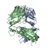
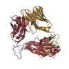
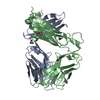
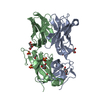
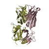
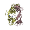
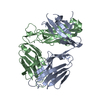
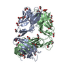
| Title | F11 T-Cell Receptor Recognising PKYVKQNTLKLAT Peptide Presented by HLA-DR*0101 | ||||||
 Components Components | (Human T-Cell Receptor ... ) x 2 ) x 2 | ||||||
 Keywords Keywords | IMMUNE SYSTEM / T Cell Receptor / Human Leukocyte Antigen / Influenza Epitope / Haemagglutinin / 3D Structure | ||||||
| Function / homology | Immunoglobulins / Immunoglobulin-like / Sandwich / Mainly Beta / DI(HYDROXYETHYL)ETHER Function and homology information Function and homology information | ||||||
| Biological species |  Homo sapiens (human) Homo sapiens (human) | ||||||
| Method | X-RAY DIFFRACTION / SYNCHROTRON / MOLECULAR REPLACEMENT / Resolution: 1.59 Å | ||||||
 Authors Authors | Rizkallah, P.J. / Cole, D.K. | ||||||
 Citation Citation | Journal: Front Immunol / Year: 2018 Title: In Silicoand Structural Analyses Demonstrate That Intrinsic Protein Motions Guide T Cell Receptor Complementarity Determining Region Loop Flexibility. Authors: Holland, C.J. / MacLachlan, B.J. / Bianchi, V. / Hesketh, S.J. / Morgan, R. / Vickery, O. / Bulek, A.M. / Fuller, A. / Godkin, A. / Sewell, A.K. / Rizkallah, P.J. / Wells, S. / Cole, D.K. | ||||||
| History |
|
- Structure visualization
| Structure viewer | Molecule: MolmilJmol/JSmol |
|---|
- Downloads & links
-Download
| PDBx/mmCIF format | 6frc.cif.gz | 204.1 KB | Display | PDBx/mmCIF format |
|---|---|---|---|---|
| PDB format | pdb6frc.ent.gz | 162.7 KB | Display | PDB format |
| PDBx/mmJSON format | 6frc.json.gz | Tree view | PDBx/mmJSON format | |
| Others |  Other downloads Other downloads |
-Validation report
| Arichive directory | https://data.pdbj.org/pub/pdb/validation_reports/fr/6frcftp://data.pdbj.org/pub/pdb/validation_reports/fr/6frc | HTTPS FTP |
|---|
-Related structure data
| Related structure data |  6eh4C  6eh5C  6eh6SC  6eh7C  6eh8C  6eh9C  6fr3C  6fr4C  6fr5C  6fr6C  6fr7C  6fr8C  6fr9C  6fraC  6frbC  6fumC 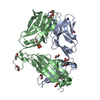 6funC  6fuoC  6fupC  6fuqC  6furC S: Starting model for refinement C: citing same article ( |
|---|---|
| Similar structure data |


-Links
 PDBj
PDBj- Assembly
| Deposited unit | 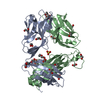
| ||||||||||||
|---|---|---|---|---|---|---|---|---|---|---|---|---|---|
| 1 |
| ||||||||||||
| Unit cell |
| ||||||||||||
| Components on special symmetry positions |
|
-Components
-Human T-Cell Receptor ... , 2 types, 2 molecules AB
| #1: Protein | Mass: 22438.043 Da / Num. of mol.: 1 Source method: isolated from a genetically manipulated source Source: (gene. exp.) Homo sapiens (human) / Production host:  Escherichia coli (E. coli) Escherichia coli (E. coli) |
|---|---|
| #2: Protein | Mass: 27208.246 Da / Num. of mol.: 1 Source method: isolated from a genetically manipulated source Source: (gene. exp.) Homo sapiens (human) / Production host: Escherichia coli (E. coli) |
-Non-polymers , 5 types, 361 molecules 


| #3: Chemical | ChemComp-EDO / Ethylene glycol Mass: 62.068 Da / Num. of mol.: 20 / Source method: obtained synthetically / Formula: C2H6O2 Mass: 62.068 Da / Num. of mol.: 20 / Source method: obtained synthetically / Formula: C2H6O2#4: Chemical | ChemComp-PEG / | Diethylene glycol Mass: 106.120 Da / Num. of mol.: 1 / Source method: obtained synthetically / Formula: C4H10O3 Mass: 106.120 Da / Num. of mol.: 1 / Source method: obtained synthetically / Formula: C4H10O3#5: Chemical | ChemComp-SO4 / | Sulfate Mass: 96.063 Da / Num. of mol.: 1 / Source method: obtained synthetically / Formula: SO4 Mass: 96.063 Da / Num. of mol.: 1 / Source method: obtained synthetically / Formula: SO4#6: Chemical | ChemComp-GOL / | Glycerol Mass: 92.094 Da / Num. of mol.: 1 / Source method: obtained synthetically / Formula: C3H8O3 Mass: 92.094 Da / Num. of mol.: 1 / Source method: obtained synthetically / Formula: C3H8O3#7: Water | ChemComp-HOH / | WaterMass: 18.015 Da / Num. of mol.: 338 / Source method: isolated from a natural source / Formula: H2O |
|---|
-Experimental details
-Experiment
| Experiment | Method: X-RAY DIFFRACTION / Number of used crystals: 1 |
|---|
- Sample preparation
| Crystal | Density Matthews: 2.5 Å3/Da / Density % sol: 50.79 % |
|---|---|
| Crystal grow | Temperature: 291 K / Method: vapor diffusion, sitting drop / pH: 6.5 / Details: 0.2M NaF, 0.1M Bis-Tris Propane, 20% PEG 3350 |
-Data collection
| Diffraction | Mean temperature: 100 K |
|---|---|
| Diffraction source | Source: SYNCHROTRON / Site: Diamond  / Beamline: I04-1 / Wavelength: 0.92 Å / Beamline: I04-1 / Wavelength: 0.92 Å |
| Detector | Type: DECTRIS PILATUS3 S 6M / Detector: PIXEL / Date: Oct 13, 2012 |
| Radiation | Protocol: SINGLE WAVELENGTH / Monochromatic (M) / Laue (L): M / Scattering type: x-ray |
| Radiation wavelength | Wavelength: 0.92 Å / Relative weight: 1 |
| Reflection | Resolution: 1.59→46.38 Å / Num. obs: 67125 / % possible obs: 99.7 % / Redundancy: 6.6 % / CC1/2: 1 / Rmerge(I) obs: 0.043 / Rrim(I) all: 0.047 / Net I/σ(I): 22.7 |
| Reflection shell | Resolution: 1.59→1.63 Å / Redundancy: 6.9 % / Rmerge(I) obs: 0.72 / Mean I/σ(I) obs: 2.9 / Num. unique obs: 4908 / CC1/2: 0.824 / Rrim(I) all: 0.779 / % possible all: 100 |
- Processing
| Software |
| ||||||||||||||||||||||||||||||||||||||||||||||||||||||||||||||||||||||||||||||||||||||||||||||||||||||||||||||||||||||||||||||||||||||||||||||||||||||||||||||||||||||||||||||||||||||
|---|---|---|---|---|---|---|---|---|---|---|---|---|---|---|---|---|---|---|---|---|---|---|---|---|---|---|---|---|---|---|---|---|---|---|---|---|---|---|---|---|---|---|---|---|---|---|---|---|---|---|---|---|---|---|---|---|---|---|---|---|---|---|---|---|---|---|---|---|---|---|---|---|---|---|---|---|---|---|---|---|---|---|---|---|---|---|---|---|---|---|---|---|---|---|---|---|---|---|---|---|---|---|---|---|---|---|---|---|---|---|---|---|---|---|---|---|---|---|---|---|---|---|---|---|---|---|---|---|---|---|---|---|---|---|---|---|---|---|---|---|---|---|---|---|---|---|---|---|---|---|---|---|---|---|---|---|---|---|---|---|---|---|---|---|---|---|---|---|---|---|---|---|---|---|---|---|---|---|---|---|---|---|---|
| Refinement | Method to determine structure: MOLECULAR REPLACEMENT Starting model: 6EH6 Resolution: 1.59→46.38 Å / Cor.coef. Fo:Fc: 0.967 / Cor.coef. Fo:Fc free: 0.958 / SU B: 3.318 / SU ML: 0.058 / Cross valid method: THROUGHOUT / ESU R: 0.083 / ESU R Free: 0.081 / Details: HYDROGENS HAVE BEEN ADDED IN THE RIDING POSITIONS
| ||||||||||||||||||||||||||||||||||||||||||||||||||||||||||||||||||||||||||||||||||||||||||||||||||||||||||||||||||||||||||||||||||||||||||||||||||||||||||||||||||||||||||||||||||||||
| Solvent computation | Ion probe radii: 0.8 Å / Shrinkage radii: 0.8 Å / VDW probe radii: 1.2 Å | ||||||||||||||||||||||||||||||||||||||||||||||||||||||||||||||||||||||||||||||||||||||||||||||||||||||||||||||||||||||||||||||||||||||||||||||||||||||||||||||||||||||||||||||||||||||
| Displacement parameters | Biso mean: 27.174 Å2
| ||||||||||||||||||||||||||||||||||||||||||||||||||||||||||||||||||||||||||||||||||||||||||||||||||||||||||||||||||||||||||||||||||||||||||||||||||||||||||||||||||||||||||||||||||||||
| Refinement step | Cycle: 1 / Resolution: 1.59→46.38 Å
| ||||||||||||||||||||||||||||||||||||||||||||||||||||||||||||||||||||||||||||||||||||||||||||||||||||||||||||||||||||||||||||||||||||||||||||||||||||||||||||||||||||||||||||||||||||||
| Refine LS restraints |
|