 Movie
Movie Controller

[English] 日本語
 Yorodumi
- PDB-2eyt: A structural basis for selection and cross-species reactivity of ... -
+ Open data

- Basic information
| Entry | Database: PDB / ID: 2eyt | ||||||
|---|---|---|---|---|---|---|---|
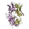
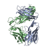
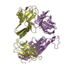
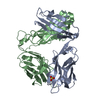
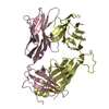
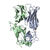
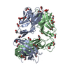
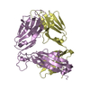
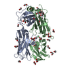
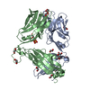
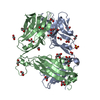
| Title | A structural basis for selection and cross-species reactivity of the semi-invariant NKT cell receptor in CD1d/glycolipid recognition | ||||||
 Components Components | (NKT15) x 2 | ||||||
 Keywords Keywords |  IMMUNE SYSTEM / Natural killer T cell receptor / NKT cell receptor / NKT15 IMMUNE SYSTEM / Natural killer T cell receptor / NKT cell receptor / NKT15 | ||||||
| Function / homology |  Function and homology information Function and homology informationalpha-beta T cell receptor complex / Translocation of ZAP-70 to Immunological synapse / Phosphorylation of CD3 and TCR zeta chains / alpha-beta T cell activation / Generation of second messenger molecules / PD-1 signaling / response to bacterium / Immunoregulatory interactions between a Lymphoid and a non-Lymphoid cell / Downstream TCR signaling / T cell receptor signaling pathway ...alpha-beta T cell receptor complex / Translocation of ZAP-70 to Immunological synapse / Phosphorylation of CD3 and TCR zeta chains / alpha-beta T cell activation / Generation of second messenger molecules / PD-1 signaling / response to bacterium / Immunoregulatory interactions between a Lymphoid and a non-Lymphoid cell / Downstream TCR signaling / T cell receptor signaling pathway / adaptive immune response / plasma membraneSimilarity search - Function | ||||||
| Biological species |  Homo sapiens (human) Homo sapiens (human) | ||||||
| Method | X-RAY DIFFRACTION / MOLECULAR REPLACEMENT / Resolution: 2.6 Å | ||||||
 Authors Authors | Kjer-Nielsen, L. / Borg, N.A. | ||||||
 Citation Citation | Journal: J.Exp.Med. / Year: 2006 Title: A structural basis for selection and cross-species reactivity of the semi-invariant NKT cell receptor in CD1d/glycolipid recognition Authors: Kjer-Nielsen, L. / Borg, N.A. / Pellicci, D.G. / Beddoe, T. / Kostenko, L. / Clements, C.S. / Williamson, N.A. / Smyth, M.J. / Besra, G.S. / Reid, H.H. / Bharadwaj, M. / Godfrey, D.I. / ...Authors: Kjer-Nielsen, L. / Borg, N.A. / Pellicci, D.G. / Beddoe, T. / Kostenko, L. / Clements, C.S. / Williamson, N.A. / Smyth, M.J. / Besra, G.S. / Reid, H.H. / Bharadwaj, M. / Godfrey, D.I. / Rossjohn, J. / McCluskey, J. | ||||||
| History |
|
- Structure visualization
| Structure viewer | Molecule: MolmilJmol/JSmol |
|---|
- Downloads & links
-Download
| PDBx/mmCIF format | 2eyt.cif.gz | 183.5 KB | Display | PDBx/mmCIF format |
|---|---|---|---|---|
| PDB format | pdb2eyt.ent.gz | 146.9 KB | Display | PDB format |
| PDBx/mmJSON format | 2eyt.json.gz | Tree view | PDBx/mmJSON format | |
| Others |  Other downloads Other downloads |
-Validation report
| Arichive directory | https://data.pdbj.org/pub/pdb/validation_reports/ey/2eytftp://data.pdbj.org/pub/pdb/validation_reports/ey/2eyt | HTTPS FTP |
|---|
-Related structure data
| Related structure data |  2eyrC  2eysC 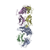 1mi5S C: citing same article ( S: Starting model for refinement |
|---|---|
| Similar structure data |


-Links
 PDBj
PDBj


- Assembly
| Deposited unit | 
| ||||||||
|---|---|---|---|---|---|---|---|---|---|
| 1 | 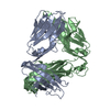
| ||||||||
| 2 | 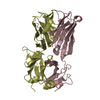
| ||||||||
| 3 | 
| ||||||||
| Unit cell |
|
-Components
| #1: Antibody | Mass: 23365.803 Da / Num. of mol.: 2 Source method: isolated from a genetically manipulated source Source: (gene. exp.) Homo sapiens (human) / Plasmid: pET30 / Species (production host): Escherichia coli / Production host:  Escherichia coli BL21(DE3) (bacteria) / Strain (production host): BL21 (DE3) / References: UniProt: Q6PIZ8, UniProt: P01848*PLUS Escherichia coli BL21(DE3) (bacteria) / Strain (production host): BL21 (DE3) / References: UniProt: Q6PIZ8, UniProt: P01848*PLUS#2: Protein | Mass: 27701.801 Da / Num. of mol.: 2 Source method: isolated from a genetically manipulated source Source: (gene. exp.) Homo sapiens (human) / Plasmid: pET30 / Species (production host): Escherichia coli / Production host: Escherichia coli BL21(DE3) (bacteria) / Strain (production host): BL21 (DE3) / References: UniProt: Q6GMR4, UniProt: A0A5B9*PLUS#3: Water | ChemComp-HOH / | Water Mass: 18.015 Da / Num. of mol.: 32 / Source method: isolated from a natural source / Formula: H2O Mass: 18.015 Da / Num. of mol.: 32 / Source method: isolated from a natural source / Formula: H2O |
|---|
-Experimental details
-Experiment
| Experiment | Method: X-RAY DIFFRACTION / Number of used crystals: 1 |
|---|
- Sample preparation
| Crystal | Density Matthews: 2.2 Å3/Da / Density % sol: 44.3 % |
|---|---|
| Crystal grow | Temperature: 297 K / Method: vapor diffusion, hanging drop / pH: 6.6 Details: 0.2M sodium sulfate, 20% PEG 3350, pH 6.6, VAPOR DIFFUSION, HANGING DROP, temperature 297K |
-Data collection
| Diffraction | Mean temperature: 100 K |
|---|---|
| Diffraction source | Source: ROTATING ANODE / Type: RIGAKU RUH3R / Wavelength: 1.5418 Å |
| Detector | Type: RIGAKU RAXIS / Detector: IMAGE PLATE / Date: Nov 7, 2004 |
| Radiation | Protocol: SINGLE WAVELENGTH / Monochromatic (M) / Laue (L): M / Scattering type: x-ray |
| Radiation wavelength | Wavelength: 1.5418 Å / Relative weight: 1 |
| Reflection | Resolution: 2.6→60.86 Å / Num. obs: 27619 / Observed criterion σ(F): 0 |
| Reflection shell | Resolution: 2.6→2.65 Å / % possible all: 98.53 |
- Processing
| Software |
| ||||||||||||||||||||||||||||||||||||||||||||||||||||||||||||||||||||||||||||||||||||||||||||||||||||||||||||||||||||||||||||||||||||||||||||||||||||||||||||||||||||||||||
|---|---|---|---|---|---|---|---|---|---|---|---|---|---|---|---|---|---|---|---|---|---|---|---|---|---|---|---|---|---|---|---|---|---|---|---|---|---|---|---|---|---|---|---|---|---|---|---|---|---|---|---|---|---|---|---|---|---|---|---|---|---|---|---|---|---|---|---|---|---|---|---|---|---|---|---|---|---|---|---|---|---|---|---|---|---|---|---|---|---|---|---|---|---|---|---|---|---|---|---|---|---|---|---|---|---|---|---|---|---|---|---|---|---|---|---|---|---|---|---|---|---|---|---|---|---|---|---|---|---|---|---|---|---|---|---|---|---|---|---|---|---|---|---|---|---|---|---|---|---|---|---|---|---|---|---|---|---|---|---|---|---|---|---|---|---|---|---|---|---|---|---|
| Refinement | Method to determine structure: MOLECULAR REPLACEMENT Starting model: PDB entry 1MI5 Resolution: 2.6→60.86 Å / Cor.coef. Fo:Fc: 0.933 / Cor.coef. Fo:Fc free: 0.897 / SU B: 30.363 / SU ML: 0.293 / Cross valid method: THROUGHOUT / σ(F): 0 / ESU R Free: 0.355 / Stereochemistry target values: MAXIMUM LIKELIHOOD / Details: HYDROGENS HAVE BEEN ADDED IN THE RIDING POSITIONS
| ||||||||||||||||||||||||||||||||||||||||||||||||||||||||||||||||||||||||||||||||||||||||||||||||||||||||||||||||||||||||||||||||||||||||||||||||||||||||||||||||||||||||||
| Solvent computation | Ion probe radii: 0.8 Å / Shrinkage radii: 0.8 Å / VDW probe radii: 1.2 Å / Solvent model: MASK | ||||||||||||||||||||||||||||||||||||||||||||||||||||||||||||||||||||||||||||||||||||||||||||||||||||||||||||||||||||||||||||||||||||||||||||||||||||||||||||||||||||||||||
| Displacement parameters | Biso mean: 42.008 Å2
| ||||||||||||||||||||||||||||||||||||||||||||||||||||||||||||||||||||||||||||||||||||||||||||||||||||||||||||||||||||||||||||||||||||||||||||||||||||||||||||||||||||||||||
| Refinement step | Cycle: LAST / Resolution: 2.6→60.86 Å
| ||||||||||||||||||||||||||||||||||||||||||||||||||||||||||||||||||||||||||||||||||||||||||||||||||||||||||||||||||||||||||||||||||||||||||||||||||||||||||||||||||||||||||
| Refine LS restraints |
| ||||||||||||||||||||||||||||||||||||||||||||||||||||||||||||||||||||||||||||||||||||||||||||||||||||||||||||||||||||||||||||||||||||||||||||||||||||||||||||||||||||||||||
| LS refinement shell | Resolution: 2.6→2.667 Å / Total num. of bins used: 20
|
