 Movie
Movie Controller


+ Open data

- Basic information
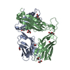
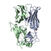
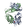
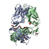
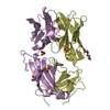
| Entry | Database: PDB / ID: 6c68 | ||||||
|---|---|---|---|---|---|---|---|
| Title | MHC-independent t cell receptor A11 | ||||||
 Components Components |
| ||||||
 Keywords Keywords |  IMMUNE SYSTEM / MHC-independent t cell receptor IMMUNE SYSTEM / MHC-independent t cell receptor | ||||||
| Function / homology | Immunoglobulins / Immunoglobulin-like / Sandwich / Mainly Beta Function and homology information Function and homology information | ||||||
| Biological species |  Mus musculus (house mouse) Mus musculus (house mouse) | ||||||
| Method | X-RAY DIFFRACTION / SYNCHROTRON / MOLECULAR REPLACEMENT / Resolution: 2.59 Å | ||||||
 Authors Authors | Lu, J. / Van Laethem, F. / Saba, I. / Chu, J. / Bhattacharya, A. / Love, N.C. / Tikhonova, A. / Radaev, S. / Sun, X. / Ko, A. ...Lu, J. / Van Laethem, F. / Saba, I. / Chu, J. / Bhattacharya, A. / Love, N.C. / Tikhonova, A. / Radaev, S. / Sun, X. / Ko, A. / Arnon, T. / Shifrut, E. / Friedman, N. / Weng, N. / Singer, A. / Sun, P.D. | ||||||
 Citation Citation | Journal: J Immunol. / Year: 2020 Title: Structure of MHC-Independent TCRs and Their Recognition of Native Antigen CD155. Authors: Lu, J. / Van Laethem, F. / Saba, I. / Chu, J. / Tikhonova, A.N. / Bhattacharya, A. / Singer, A. / Sun, P.D. | ||||||
| History |
|
- Structure visualization
| Structure viewer | Molecule: MolmilJmol/JSmol |
|---|
- Downloads & links
-Download
| PDBx/mmCIF format | 6c68.cif.gz | 186.3 KB | Display | PDBx/mmCIF format |
|---|---|---|---|---|
| PDB format | pdb6c68.ent.gz | 147.8 KB | Display | PDB format |
| PDBx/mmJSON format | 6c68.json.gz | Tree view | PDBx/mmJSON format | |
| Others |  Other downloads Other downloads |
-Validation report
| Arichive directory | https://data.pdbj.org/pub/pdb/validation_reports/c6/6c68ftp://data.pdbj.org/pub/pdb/validation_reports/c6/6c68 | HTTPS FTP |
|---|
-Related structure data
| Related structure data |  6c61C 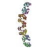 3c5zS S: Starting model for refinement C: citing same article ( |
|---|---|
| Similar structure data |


-Links
 PDBj
PDBj- Assembly
| Deposited unit | 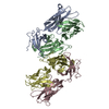
| ||||||||
|---|---|---|---|---|---|---|---|---|---|
| 1 | 
| ||||||||
| 2 | 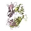
| ||||||||
| Unit cell |
|
-Components
| #1: Protein | Mass: 23063.623 Da / Num. of mol.: 2 Source method: isolated from a genetically manipulated source Source: (gene. exp.) Mus musculus (house mouse) / Production host:  Escherichia coli (E. coli) Escherichia coli (E. coli)#2: Protein | Mass: 27047.943 Da / Num. of mol.: 2 Source method: isolated from a genetically manipulated source Source: (gene. exp.) Mus musculus (house mouse) / Production host: Escherichia coli (E. coli)#3: Water | ChemComp-HOH / | Water Mass: 18.015 Da / Num. of mol.: 140 / Source method: isolated from a natural source / Formula: H2O Mass: 18.015 Da / Num. of mol.: 140 / Source method: isolated from a natural source / Formula: H2O |
|---|
-Experimental details
-Experiment
| Experiment | Method: X-RAY DIFFRACTION / Number of used crystals: 1 |
|---|
- Sample preparation
| Crystal | Density Matthews: 2.71 Å3/Da / Density % sol: 54.55 % |
|---|---|
| Crystal grow | Temperature: 293 K / Method: vapor diffusion, hanging drop / Details: 20% PEG 4000, 0.1M Tris, pH8.5, 10mM MgCl2 |
-Data collection
| Diffraction | Mean temperature: 100 K |
|---|---|
| Diffraction source | Source: SYNCHROTRON / Site: APS  / Beamline: 22-ID / Wavelength: 1 Å / Beamline: 22-ID / Wavelength: 1 Å |
| Detector | Type: MAR CCD 165 mm / Detector: CCD / Date: Nov 18, 2011 |
| Radiation | Protocol: SINGLE WAVELENGTH / Monochromatic (M) / Laue (L): M / Scattering type: x-ray |
| Radiation wavelength | Wavelength: 1 Å / Relative weight: 1 |
| Reflection | Resolution: 2.6→50 Å / Num. obs: 32187 / % possible obs: 94.7 % / Observed criterion σ(I): 2.1 / Redundancy: 9.9 % / Biso Wilson estimate: 69.46 Å2 / Rmerge(I) obs: 0.095 / Net I/σ(I): 23 |
| Reflection shell | Resolution: 2.6→2.65 Å / Redundancy: 4 % / Rmerge(I) obs: 0.487 / Num. unique obs: 1262 / % possible all: 68.4 |
- Processing
| Software |
| ||||||||||||||||||||||||||||||||||||||||||||||||||||||||||||||||||||||||||||||||||||||||||||||||||||||||||||||||||
|---|---|---|---|---|---|---|---|---|---|---|---|---|---|---|---|---|---|---|---|---|---|---|---|---|---|---|---|---|---|---|---|---|---|---|---|---|---|---|---|---|---|---|---|---|---|---|---|---|---|---|---|---|---|---|---|---|---|---|---|---|---|---|---|---|---|---|---|---|---|---|---|---|---|---|---|---|---|---|---|---|---|---|---|---|---|---|---|---|---|---|---|---|---|---|---|---|---|---|---|---|---|---|---|---|---|---|---|---|---|---|---|---|---|---|---|
| Refinement | Method to determine structure: MOLECULAR REPLACEMENT Starting model: 3c5Z Resolution: 2.59→34.02 Å / Cor.coef. Fo:Fc: 0.9237 / Cor.coef. Fo:Fc free: 0.8898 / Cross valid method: THROUGHOUT / σ(F): 0
| ||||||||||||||||||||||||||||||||||||||||||||||||||||||||||||||||||||||||||||||||||||||||||||||||||||||||||||||||||
| Displacement parameters | Biso mean: 71.58 Å2
| ||||||||||||||||||||||||||||||||||||||||||||||||||||||||||||||||||||||||||||||||||||||||||||||||||||||||||||||||||
| Refine analyze | Luzzati coordinate error obs: 0.361 Å | ||||||||||||||||||||||||||||||||||||||||||||||||||||||||||||||||||||||||||||||||||||||||||||||||||||||||||||||||||
| Refinement step | Cycle: 1 / Resolution: 2.59→34.02 Å
| ||||||||||||||||||||||||||||||||||||||||||||||||||||||||||||||||||||||||||||||||||||||||||||||||||||||||||||||||||
| Refine LS restraints |
| ||||||||||||||||||||||||||||||||||||||||||||||||||||||||||||||||||||||||||||||||||||||||||||||||||||||||||||||||||
| LS refinement shell | Resolution: 2.59→2.67 Å / Total num. of bins used: 16
|
