phosphopantetheine binding / 3-oxoacyl-[acyl-carrier-protein] synthase activity / Transferases; Acyltransferases; Transferring groups other than aminoacyl groups / fatty acid biosynthetic process Similarity search - Function
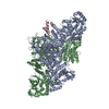
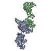
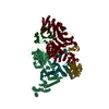
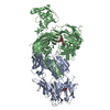
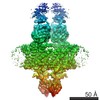
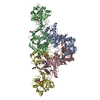
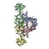
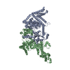
Journal: Nat Chem Biol / Year: 2018 Title: The structural organization of substrate loading in iterative polyketide synthases. Authors: Dominik A Herbst / Callie R Huitt-Roehl / Roman P Jakob / Jacob M Kravetz / Philip A Storm / Jamie R Alley / Craig A Townsend / Timm Maier / Abstract: Polyketide synthases (PKSs) are microbial multienzymes for the biosynthesis of biologically potent secondary metabolites. Polyketide production is initiated by the loading of a starter unit onto an ...Polyketide synthases (PKSs) are microbial multienzymes for the biosynthesis of biologically potent secondary metabolites. Polyketide production is initiated by the loading of a starter unit onto an integral acyl carrier protein (ACP) and its subsequent transfer to the ketosynthase (KS). Initial substrate loading is achieved either by multidomain loading modules or by the integration of designated loading domains, such as starter unit acyltransferases (SAT), whose structural integration into PKS remains unresolved. A crystal structure of the loading/condensing region of the nonreducing PKS CTB1 demonstrates the ordered insertion of a pseudodimeric SAT into the condensing region, which is aided by the SAT-KS linker. Cryo-electron microscopy of the post-loading state trapped by mechanism-based crosslinking of ACP to KS reveals asymmetry across the CTB1 loading/-condensing region, in accord with preferential 1:2 binding stoichiometry. These results are critical for re-engineering the loading step in polyketide biosynthesis and support functional relevance of asymmetric conformations of PKSs.
Resolution: 2.77→63.45 Å / SU ML: 0.46 / Cross valid method: FREE R-VALUE / σ(F): 1.34 / Phase error: 27.1 / Stereochemistry target values: ML Details: UNIDENTIFIED DIFFERENCE DENSITY AROUND THE ACTIVE SITE RESIDUES C553 AND C119, POSSIBLY DUE TO PARTIAL ACYLATION OR OXIDATION. THE DIFFERENCE DENSITY IN THE REGION AROUND RESIDUES 651 TO 661 ...Details: UNIDENTIFIED DIFFERENCE DENSITY AROUND THE ACTIVE SITE RESIDUES C553 AND C119, POSSIBLY DUE TO PARTIAL ACYLATION OR OXIDATION. THE DIFFERENCE DENSITY IN THE REGION AROUND RESIDUES 651 TO 661 INDICATES MULTIPLE DIFFERENT MAIN CHAIN CONFORMATION, WHEREAS ONLY ONE COULD ME MODELLED. BASED ON OBSERVED B-FACTORS AND THE FACT THAT CRYSTALLIZATION WAS CARRIED OUT IN THE PRESENCE OF 0.3M MG(2+) SOME SURFACE WATERS MIGHT ALSO REPRESENT PARTIALLY OCCUPIED HYDRATED MAGNESIUM IONS.
Rfactor
Num. reflection
% reflection
Rfree
0.2405
3916
4.91 %
Rwork
0.2052
-
-
obs
0.2069
79812
99.04 %
Solvent computation
Shrinkage radii: 0.9 Å / VDW probe radii: 1.11 Å / Solvent model: FLAT BULK SOLVENT MODEL
Refinement step
Cycle: LAST / Resolution: 2.77→63.45 Å
Protein
Nucleic acid
Ligand
Solvent
Total
Num. atoms
19291
0
69
352
19712
Refine LS restraints
Refine-ID
Type
Dev ideal
Number
X-RAY DIFFRACTION
f_bond_d
0.002
19940
X-RAY DIFFRACTION
f_angle_d
0.496
27153
X-RAY DIFFRACTION
f_dihedral_angle_d
11.925
12022
X-RAY DIFFRACTION
f_chiral_restr
0.04
3085
X-RAY DIFFRACTION
f_plane_restr
0.003
3570
LS refinement shell
Resolution (Å)
Rfactor Rfree
Num. reflection Rfree
Rfactor Rwork
Num. reflection Rwork
Refine-ID
% reflection obs (%)
2.7698-2.8036
0.4542
132
0.4626
2366
X-RAY DIFFRACTION
89
2.8036-2.8391
0.4397
127
0.4051
2551
X-RAY DIFFRACTION
93
2.8391-2.8764
0.3767
125
0.3673
2608
X-RAY DIFFRACTION
96
2.8764-2.9158
0.366
129
0.3536
2699
X-RAY DIFFRACTION
99
2.9158-2.9575
0.386
125
0.322
2707
X-RAY DIFFRACTION
99
2.9575-3.0016
0.3045
143
0.3039
2673
X-RAY DIFFRACTION
100
3.0016-3.0485
0.3249
140
0.2864
2698
X-RAY DIFFRACTION
100
3.0485-3.0985
0.3333
141
0.2847
2742
X-RAY DIFFRACTION
100
3.0985-3.1519
0.3209
142
0.2757
2694
X-RAY DIFFRACTION
100
3.1519-3.2092
0.3512
144
0.269
2705
X-RAY DIFFRACTION
100
3.2092-3.271
0.3081
142
0.258
2721
X-RAY DIFFRACTION
100
3.271-3.3377
0.3009
140
0.2538
2688
X-RAY DIFFRACTION
100
3.3377-3.4103
0.2856
122
0.2445
2721
X-RAY DIFFRACTION
100
3.4103-3.4896
0.2552
145
0.2282
2754
X-RAY DIFFRACTION
100
3.4896-3.5769
0.235
145
0.2149
2686
X-RAY DIFFRACTION
100
3.5769-3.6736
0.2382
153
0.2119
2714
X-RAY DIFFRACTION
100
3.6736-3.7817
0.2397
154
0.1903
2719
X-RAY DIFFRACTION
100
3.7817-3.9037
0.1993
131
0.1935
2732
X-RAY DIFFRACTION
100
3.9037-4.0432
0.2272
137
0.1779
2732
X-RAY DIFFRACTION
100
4.0432-4.2051
0.2067
145
0.1749
2731
X-RAY DIFFRACTION
100
4.2051-4.3964
0.2364
154
0.1549
2730
X-RAY DIFFRACTION
100
4.3964-4.6281
0.1814
138
0.1529
2746
X-RAY DIFFRACTION
100
4.6281-4.918
0.1718
149
0.149
2756
X-RAY DIFFRACTION
100
4.918-5.2975
0.1776
135
0.16
2761
X-RAY DIFFRACTION
100
5.2975-5.8303
0.2156
150
0.1756
2749
X-RAY DIFFRACTION
100
5.8303-6.6732
0.2107
144
0.1842
2796
X-RAY DIFFRACTION
100
6.6732-8.4044
0.2003
136
0.1675
2814
X-RAY DIFFRACTION
100
8.4044-63.4666
0.2309
148
0.19
2903
X-RAY DIFFRACTION
99
Refinement TLS params.
Method: refined / Refine-ID: X-RAY DIFFRACTION
ID
L11 (°2)
L12 (°2)
L13 (°2)
L22 (°2)
L23 (°2)
L33 (°2)
S11 (Å °)
S12 (Å °)
S13 (Å °)
S21 (Å °)
S22 (Å °)
S23 (Å °)
S31 (Å °)
S32 (Å °)
S33 (Å °)
T11 (Å2)
T12 (Å2)
T13 (Å2)
T22 (Å2)
T23 (Å2)
T33 (Å2)
Origin x (Å)
Origin y (Å)
Origin z (Å)
1
0.2305
0.0516
0.068
2.5314
-0.8064
0.5756
0.0381
-0.0539
0.1342
0.0927
-0.0126
0.3934
-0.0409
-0.027
-0.0428
0.5078
-0.1181
0.0021
0.5741
-0.0819
0.5185
-6.2681
85.6464
-22.2192
2
1.7655
0.8284
0.2215
2.6664
0.3566
1.3516
0.2133
-0.2788
0.3746
0.4609
-0.1684
0.1906
-0.1141
0.1333
-0.0477
0.4688
-0.1059
0.0219
0.4324
-0.0098
0.3535
-2.7195
48.3417
-7.6392
3
2.3819
0.6164
1.2024
0.5422
0.1984
1.1419
0.1742
-0.2866
0.0555
0.0945
-0.1231
0.2464
0.1814
-0.2935
-0.0494
0.4609
-0.0506
-0.0134
0.4389
0.0129
0.5061
-43.3764
26.2176
-29.1474
4
2.0559
1.0196
0.641
1.9846
0.9359
2.2877
-0.0781
0.0811
0.2811
-0.0072
0.0838
-0.0026
-0.2988
0.2446
-0.0179
0.4555
-0.0457
-0.1665
0.4418
0.0383
0.6883
-33.0831
58.356
-50.3213
5
1.2551
0.3404
0.3419
1.0977
0.1774
0.9035
0.0247
0.1637
-0.0968
-0.114
0.0233
-0.0545
0.0847
0.0276
-0.0589
0.3357
-0.0063
-0.0295
0.4102
-0.0072
0.2687
10.8736
46.3087
-35.6
6
0.4819
0.2544
0.1324
3.0008
0.2571
0.1008
-0.0442
0.0952
0.1029
0.2035
0.0265
-0.6904
-0.119
0.2067
0.0127
0.5035
-0.0987
-0.0989
0.5918
0.0682
0.5757
34.3809
91.4807
-25.4051
Refinement TLS group
ID
Refine-ID
Refine TLS-ID
Selection details
1
X-RAY DIFFRACTION
1
chain 'A' and (resid5through441 )
2
X-RAY DIFFRACTION
2
chain 'A' and (resid442through805 )
3
X-RAY DIFFRACTION
3
chain 'A' and (resid806through1288 )
4
X-RAY DIFFRACTION
4
chain 'B' and (resid5through337 )
5
X-RAY DIFFRACTION
5
chain 'B' and (resid338through823 )
6
X-RAY DIFFRACTION
6
chain 'B' and (resid824through1288 )
+
About Yorodumi
-
News
-
Feb 9, 2022. New format data for meta-information of EMDB entries
New format data for meta-information of EMDB entries
Version 3 of the EMDB header file is now the official format.
The previous official version 1.9 will be removed from the archive.
In the structure databanks used in Yorodumi, some data are registered as the other names, "COVID-19 virus" and "2019-nCoV". Here are the details of the virus and the list of structure data.
Jan 31, 2019. EMDB accession codes are about to change! (news from PDBe EMDB page)
EMDB accession codes are about to change! (news from PDBe EMDB page)
The allocation of 4 digits for EMDB accession codes will soon come to an end. Whilst these codes will remain in use, new EMDB accession codes will include an additional digit and will expand incrementally as the available range of codes is exhausted. The current 4-digit format prefixed with “EMD-” (i.e. EMD-XXXX) will advance to a 5-digit format (i.e. EMD-XXXXX), and so on. It is currently estimated that the 4-digit codes will be depleted around Spring 2019, at which point the 5-digit format will come into force.
The EM Navigator/Yorodumi systems omit the EMD- prefix.
Related info.:Q: What is EMD? / ID/Accession-code notation in Yorodumi/EM Navigator
Yorodumi is a browser for structure data from EMDB, PDB, SASBDB, etc.
This page is also the successor to EM Navigator detail page, and also detail information page/front-end page for Omokage search.
The word "yorodu" (or yorozu) is an old Japanese word meaning "ten thousand". "mi" (miru) is to see.
Related info.:EMDB / PDB / SASBDB / Comparison of 3 databanks / Yorodumi Search / Aug 31, 2016. New EM Navigator & Yorodumi / Yorodumi Papers / Jmol/JSmol / Function and homology information / Changes in new EM Navigator and Yorodumi
 Movie
Movie Controller

 Yorodumi
 Open data
 Basic information
 Components
 Keywords
 Function and homology information
 Authors
 Switzerland,
  United States, 5items
  Citation
 Structure visualization
 Downloads & links
 Other downloads
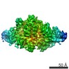


 PDBj
PDBj


 Assembly
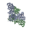


 Mass: 62.068 Da / Num. of mol.: 12 / Source method: obtained synthetically / Formula: C2H6O2
 Mass: 92.094 Da / Num. of mol.: 2 / Source method: obtained synthetically / Formula: C3H8O3
 Mass: 24.305 Da / Num. of mol.: 1 / Source method: obtained synthetically / Formula: Mg
 Mass: 154.251 Da / Num. of mol.: 1 / Source method: isolated from a natural source / Formula: C4H10O2S2
 Sample preparation
 Processing