[English] 日本語
Yorodumi
- PDB-4ro5: Crystal structure of the SAT domain from the non-reducing fungal ... -

+
Open data


ID or keywords:

Loading...

-
Basic information

Entry
Database: PDB / ID: 4ro5
TitleCrystal structure of the SAT domain from the non-reducing fungal polyketide synthase CazM
ComponentsSAT domain from CazM
Keywords
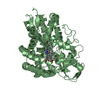
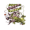
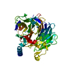
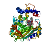
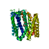
TRANSFERASE / non reducing polyketide synthase / acyl carrier protein transacylase
Function / homology
Function and homology information


3-oxoacyl-[acyl-carrier-protein] synthase activity / fatty acid biosynthetic process
Similarity search - Function
Starter unit:ACP transacylase / Starter unit:ACP transacylase in aflatoxin biosynthesis / Malonyl-CoA ACP transacylase, ACP-binding / Acyl transferase domain superfamily / Acyl transferase / Acyl transferase domain / Acyl transferase domain in polyketide synthase (PKS) enzymes. / Acyl transferase/acyl hydrolase/lysophospholipase / Ketosynthase family 3 (KS3) domain profile. / Beta-ketoacyl synthase ...Starter unit:ACP transacylase / Starter unit:ACP transacylase in aflatoxin biosynthesis / Malonyl-CoA ACP transacylase, ACP-binding / Acyl transferase domain superfamily / Acyl transferase / Acyl transferase domain / Acyl transferase domain in polyketide synthase (PKS) enzymes. / Acyl transferase/acyl hydrolase/lysophospholipase / Ketosynthase family 3 (KS3) domain profile. / Beta-ketoacyl synthase / Beta-ketoacyl synthase, active site / Ketosynthase family 3 (KS3) active site signature. / Polyketide synthase, beta-ketoacyl synthase domain / Beta-ketoacyl synthase, N-terminal / Beta-ketoacyl synthase, C-terminal / Beta-ketoacyl synthase, N-terminal domain / Beta-ketoacyl synthase, C-terminal domain / Thiolase-like
Similarity search - Domain/homology
Ketosynthase family 3 (KS3) domain-containing protein
Similarity search - Component
Biological speciesChaetomium globosum CBS 148.51 (fungus)
MethodX-RAY DIFFRACTION / SYNCHROTRON / SAD / Resolution: 1.6 Å
AuthorsWinter, J.M. / Cascio, D. / Sawaya, M.R. / Tang, Y.
CitationJournal: J.Am.Chem.Soc. / Year: 2015
Title: Biochemical and Structural Basis for Controlling Chemical Modularity in Fungal Polyketide Biosynthesis.
Authors: Winter, J.M. / Cascio, D. / Dietrich, D. / Sato, M. / Watanabe, K. / Sawaya, M.R. / Vederas, J.C. / Tang, Y.
History
DepositionOct 27, 2014Deposition site: RCSB / Processing site: RCSB
Revision 1.0Sep 9, 2015Provider: repository / Type: Initial release
Revision 1.1Nov 22, 2017Group: Refinement description / Category: software

-
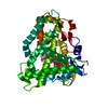
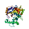
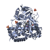
Structure visualization

Structure viewerMolecule:
MolmilJmol/JSmol

Downloads & links

-
Assembly

Deposited unit
A: SAT domain from CazM
hetero molecules


Theoretical massNumber of molelcules
Total (without water)44,7603
Polymers44,5761
Non-polymers1842
Water3,225179
1


  • Idetical with deposited unit
  • defined by author&software
TypeNameSymmetry operationNumber
identity operation1_555x,y,z1
MethodPISA
Unit cell
Length a, b, c (Å)43.130, 52.160, 163.090
Angle α, β, γ (deg.)90.000, 90.000, 90.000
Int Tables number19
Space group name H-MP212121

-
Components

#1: Protein SAT domain from CazM


Mass: 44576.082 Da / Num. of mol.: 1
Source method: isolated from a genetically manipulated source
Source: (gene. exp.) Chaetomium globosum CBS 148.51 (fungus)
Strain: CBS 148.51 / Gene: cazM, CHGG_07645 / Plasmid: pHis8 / Production host: Escherichia coli (E. coli) / Strain (production host): BL21(DE3) / References: UniProt: Q2GWK9
#2: Chemical ChemComp-GOL / GLYCEROL / GLYCERIN / PROPANE-1,2,3-TRIOL / Glycerol


Mass: 92.094 Da / Num. of mol.: 2 / Source method: obtained synthetically / Formula: C3H8O3
#3: Water ChemComp-HOH / water / Water


Mass: 18.015 Da / Num. of mol.: 179 / Source method: isolated from a natural source / Formula: H2O
Sequence detailsAS PER THE AUTH THE SEQUENCE REPRESENTED BY UNP Q2GWK9 IS INCORRECT IN THIS REGION. THE AUTHORS ...AS PER THE AUTH THE SEQUENCE REPRESENTED BY UNP Q2GWK9 IS INCORRECT IN THIS REGION. THE AUTHORS RESEQUENCED THIS REGION AND FOUND AN EXTRA BASE CAUSING A DISCREPANCY BETWEEN THE AUTHORS SEQUENCE AND UNIPROT SEQ. THIS DISCREPANCY CAUSES THE ARTIFICIAL OCCURRENCE OF AN EXTRA INTRON IN THAT REGION. THIS ENTIRE REGION WAS RE-SEQUENCED BY THE AUTHORS USING CDNA AND THEY HAVE AN ACTIVE ENZYME AND THEIR SEQUENCE MATCHES TO THE 4RO5 CRYSTAL STRUCTURE AT 1.6A RESOLUTION. THE AUTHORS WILL DEPOSIT THE CORRECTED SEQUENCE SOON

-
Experimental details

-
Experiment

ExperimentMethod: X-RAY DIFFRACTION / Number of used crystals: 1

-
Sample preparation

CrystalDensity Matthews: 2.06 Å3/Da / Density % sol: 40.29 %
Crystal growTemperature: 277 K / Method: vapor diffusion, hanging drop / pH: 7
Details: 100 mM Tris HCl pH 7 and 20% PEG 8000, vapor diffusion, hanging drop, temperature 277K

-
Data collection

DiffractionMean temperature: 100 K
Diffraction sourceSource: SYNCHROTRON / Site: APS / Beamline: 24-ID-C / Wavelength: 0.942 Å
DetectorType: DECTRIS PILATUS 6M / Detector: PIXEL / Date: Dec 8, 2013
RadiationProtocol: SINGLE WAVELENGTH / Monochromatic (M) / Laue (L): M / Scattering type: x-ray
Radiation wavelengthWavelength: 0.942 Å / Relative weight: 1
ReflectionResolution: 1.6→81.54 Å / Num. all: 87226 / Num. obs: 48603 / % possible obs: 92.7 % / Observed criterion σ(I): -3 / Redundancy: 1.8 % / Biso Wilson estimate: 12.8 Å2 / Rmerge(I) obs: 0.047 / Rsym value: 0.047 / Χ2: 1.963 / Net I/σ(I): 11.53
Reflection shell
Resolution (Å)Rmerge(I) obsMean I/σ(I) obsNum. measured obsNum. unique obsDiffraction-ID% possible all
1.6-1.640.2642.98106686112188.1
1.64-1.680.2363.42116216501195.4
1.68-1.730.2034.05112396278194.5
1.73-1.790.1624.85105876017193.8
1.79-1.850.1355.6498005685192.7
1.85-1.910.1176.9104595710195.2
1.91-1.980.0968.4101015559194.8
1.98-2.060.07810.194955293195.1
2.06-2.160.06611.7885794867190.9
2.16-2.260.05813.3883544788193.1
2.26-2.380.05215.1183434550194.5
2.38-2.530.04815.8378634307193.2
2.53-2.70.04417.471934010192.5
2.7-2.920.03918.8763393598190.1
2.92-3.20.03221.8665063454193.4
3.2-3.570.0323.9256313024190.2
3.57-4.130.02625.1745962567187.4
4.13-5.050.02526.9943142258189.9
5.05-7.150.02525.6733301724189
7.15-81.540.02127.881765924186.7

-
Processing

Software
NameVersionClassificationNB
XSCALEdata scaling
PHENIXrefinement
PDB_EXTRACT3.15data extraction
XDSdata reduction
SHELXCDphasing
SHELXEmodel building
RefinementMethod to determine structure: SAD / Resolution: 1.6→81.54 Å / SU ML: 0.16 / Cross valid method: THROUGHOUT / σ(F): 0 / Phase error: 19.23 / Stereochemistry target values: ML
RfactorNum. reflection% reflectionSelection details
Rfree0.2153 2455 5.05 %RANDOM
Rwork0.187 ---
obs0.1884 48603 97.75 %-
all-48603 --
Solvent computationShrinkage radii: 0.9 Å / VDW probe radii: 1.11 Å / Solvent model: FLAT BULK SOLVENT MODEL
Displacement parametersBiso max: 56.31 Å2 / Biso mean: 15.6292 Å2 / Biso min: 6.04 Å2
Refinement stepCycle: LAST / Resolution: 1.6→81.54 Å
ProteinNucleic acidLigandSolventTotal
Num. atoms2980 0 12 179 3171
Refine LS restraints
Refine-IDTypeDev idealNumber
X-RAY DIFFRACTIONf_bond_d0.0063054
X-RAY DIFFRACTIONf_angle_d1.0594161
X-RAY DIFFRACTIONf_chiral_restr0.041476
X-RAY DIFFRACTIONf_plane_restr0.005546
X-RAY DIFFRACTIONf_dihedral_angle_d11.441113
LS refinement shell

Refine-ID: X-RAY DIFFRACTION / Total num. of bins used: 18

Resolution (Å)Rfactor RfreeNum. reflection RfreeRfactor RworkNum. reflection RworkNum. reflection all% reflection obs (%)
1.6-1.62920.25171180.22204232286
1.6292-1.66240.24521390.18842433257294
1.6624-1.69860.18991260.17812485261196
1.6986-1.73810.23551200.17812525264596
1.7381-1.78160.21631290.16842445257496
1.7816-1.82970.24041300.18082465259596
1.8297-1.88360.23281320.18072574270698
1.8836-1.94440.20751290.18542577270699
1.9444-2.01390.20781380.17212570270899
2.0139-2.09450.1971330.17252592272599
2.0945-2.18980.21171490.183825962745100
2.1898-2.30530.18621400.180326002740100
2.3053-2.44970.22971440.185326182762100
2.4497-2.63890.23771490.190226162765100
2.6389-2.90450.23041360.200126632799100
2.9045-3.32480.19761430.202726542797100
3.3248-4.18890.2221420.183926942836100
4.1889-81.540.20241580.192428372995100
Refinement TLS params.Method: refined / Origin x: 28.6139 Å / Origin y: 27.886 Å / Origin z: 19.3474 Å
111213212223313233
T0.071 Å2-0.0083 Å2-0.0007 Å2-0.0833 Å20.0057 Å2--0.0834 Å2
L0.436 °2-0.2178 °2-0.0378 °2-0.5578 °20.0583 °2--0.267 °2
S0.0095 Å °0.009 Å °0.0409 Å °0.0076 Å °-0.0083 Å °-0.0502 Å °-0.0351 Å °0.0152 Å °-0.0004 Å °
Refinement TLS group
IDRefine-IDRefine TLS-IDSelection detailsAuth asym-IDAuth seq-ID
1X-RAY DIFFRACTION1allA1 - 396
2X-RAY DIFFRACTION1allA1 - 679
3X-RAY DIFFRACTION1allA1 - 401
4X-RAY DIFFRACTION1allA2 - 402

+
About Yorodumi

-
News

-
Feb 9, 2022. New format data for meta-information of EMDB entries

New format data for meta-information of EMDB entries

  • Version 3 of the EMDB header file is now the official format.
  • The previous official version 1.9 will be removed from the archive.

Related info.:EMDB header

External links:wwPDB to switch to version 3 of the EMDB data model

-
Aug 12, 2020. Covid-19 info

Covid-19 info

URL: https://pdbj.org/emnavi/covid19.php

New page: Covid-19 featured information page in EM Navigator.

Related info.:Covid-19 info / Mar 5, 2020. Novel coronavirus structure data

+
Mar 5, 2020. Novel coronavirus structure data

Novel coronavirus structure data

Related info.:Yorodumi Speices / Aug 12, 2020. Covid-19 info

External links:COVID-19 featured content - PDBj / Molecule of the Month (242):Coronavirus Proteases

+
Jan 31, 2019. EMDB accession codes are about to change! (news from PDBe EMDB page)

EMDB accession codes are about to change! (news from PDBe EMDB page)

  • The allocation of 4 digits for EMDB accession codes will soon come to an end. Whilst these codes will remain in use, new EMDB accession codes will include an additional digit and will expand incrementally as the available range of codes is exhausted. The current 4-digit format prefixed with “EMD-” (i.e. EMD-XXXX) will advance to a 5-digit format (i.e. EMD-XXXXX), and so on. It is currently estimated that the 4-digit codes will be depleted around Spring 2019, at which point the 5-digit format will come into force.
  • The EM Navigator/Yorodumi systems omit the EMD- prefix.

Related info.:Q: What is EMD? / ID/Accession-code notation in Yorodumi/EM Navigator

External links:EMDB Accession Codes are Changing Soon! / Contact to PDBj

+
Jul 12, 2017. Major update of PDB

Major update of PDB

  • wwPDB released updated PDB data conforming to the new PDBx/mmCIF dictionary.
  • This is a major update changing the version number from 4 to 5, and with Remediation, in which all the entries are updated.
  • In this update, many items about electron microscopy experimental information are reorganized (e.g. em_software).
  • Now, EM Navigator and Yorodumi are based on the updated data.

External links:wwPDB Remediation / Enriched Model Files Conforming to OneDep Data Standards Now Available in the PDB FTP Archive

-
Yorodumi

Thousand views of thousand structures

  • Yorodumi is a browser for structure data from EMDB, PDB, SASBDB, etc.
  • This page is also the successor to EM Navigator detail page, and also detail information page/front-end page for Omokage search.
  • The word "yorodu" (or yorozu) is an old Japanese word meaning "ten thousand". "mi" (miru) is to see.

Related info.:EMDB / PDB / SASBDB / Comparison of 3 databanks / Yorodumi Search / Aug 31, 2016. New EM Navigator & Yorodumi / Yorodumi Papers / Jmol/JSmol / Function and homology information / Changes in new EM Navigator and Yorodumi

Read more