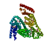
 Movie
Movie Controller


+ Open data

- Basic information
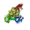
| Entry | Database: PDB / ID: 6ezq | ||||||
|---|---|---|---|---|---|---|---|
| Title | human Serum Albumin complexed with NBD-C12 fatty acid | ||||||
 Components Components | Serum albumin | ||||||
 Keywords Keywords | TRANSPORT PROTEIN / fatty acid binding site human Serum Albumin NBD Label drug interaction | ||||||
| Function / homology |  Function and homology information Function and homology informationcellular response to calcium ion starvation / exogenous protein binding / Ciprofloxacin ADME / enterobactin binding / Heme biosynthesis / HDL remodeling / negative regulation of mitochondrial depolarization / Prednisone ADME / Heme degradation / Aspirin ADME ...cellular response to calcium ion starvation / exogenous protein binding / Ciprofloxacin ADME / enterobactin binding / Heme biosynthesis / HDL remodeling / negative regulation of mitochondrial depolarization / Prednisone ADME / Heme degradation / Aspirin ADME / antioxidant activity / toxic substance binding / small molecule binding / Scavenging of heme from plasma / Recycling of bile acids and salts / cellular response to starvation / platelet alpha granule lumen / fatty acid binding / Post-translational protein phosphorylation / Cytoprotection by HMOX1 / Regulation of Insulin-like Growth Factor (IGF) transport and uptake by Insulin-like Growth Factor Binding Proteins (IGFBPs) / pyridoxal phosphate binding / Platelet degranulation / protein-folding chaperone binding / blood microparticle / copper ion binding / endoplasmic reticulum lumen / Golgi apparatus / endoplasmic reticulum / protein-containing complex / DNA binding / extracellular space / extracellular exosome / extracellular region / identical protein binding / nucleus / cytoplasmSimilarity search - Function | ||||||
| Biological species |  Homo sapiens (human) Homo sapiens (human) | ||||||
| Method | X-RAY DIFFRACTION / SYNCHROTRON / Resolution: 2.39 Å | ||||||
 Authors Authors | Wenskowsky, L. / Liesum, A. / Schreuder, H.A. | ||||||
 Citation Citation | Journal: Angew. Chem. Int. Ed. Engl. / Year: 2018 Title: Identification and Characterization of a Single High-Affinity Fatty Acid Binding Site in Human Serum Albumin. Authors: Wenskowsky, L. / Schreuder, H. / Derdau, V. / Matter, H. / Volkmar, J. / Nazare, M. / Opatz, T. / Petry, S. | ||||||
| History |
|
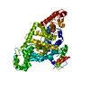
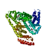
- Structure visualization
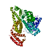
| Structure viewer | Molecule: MolmilJmol/JSmol |
|---|
- Downloads & links
-Download
| PDBx/mmCIF format | 6ezq.cif.gz | 129.9 KB | Display | PDBx/mmCIF format |
|---|---|---|---|---|
| PDB format | pdb6ezq.ent.gz | 104.5 KB | Display | PDB format |
| PDBx/mmJSON format | 6ezq.json.gz | Tree view | PDBx/mmJSON format | |
| Others |  Other downloads Other downloads |
-Validation report
| Arichive directory | https://data.pdbj.org/pub/pdb/validation_reports/ez/6ezqftp://data.pdbj.org/pub/pdb/validation_reports/ez/6ezq | HTTPS FTP |
|---|
-Related structure data
| Similar structure data |
|---|


-Links
 PDBj
PDBj


- Assembly
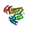
| Deposited unit | 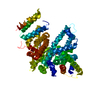
| ||||||||
|---|---|---|---|---|---|---|---|---|---|
| 1 |
| ||||||||
| Unit cell |
|
-Components
| #1: Protein | Mass: 66571.219 Da / Num. of mol.: 1 / Source method: isolated from a natural source / Source: (natural) Homo sapiens (human) / Tissue: serum / References: UniProt: P02768 | ||
|---|---|---|---|
| #2: Chemical |   Mass: 378.423 Da / Num. of mol.: 2 / Source method: obtained synthetically / Formula: C18H26N4O5 Mass: 378.423 Da / Num. of mol.: 2 / Source method: obtained synthetically / Formula: C18H26N4O5#3: Water | ChemComp-HOH / | Water Mass: 18.015 Da / Num. of mol.: 324 / Source method: isolated from a natural source / Formula: H2O Mass: 18.015 Da / Num. of mol.: 324 / Source method: isolated from a natural source / Formula: H2O |
-Experimental details
-Experiment
| Experiment | Method: X-RAY DIFFRACTION / Number of used crystals: 1 |
|---|
- Sample preparation
| Crystal | Density Matthews: 2.35 Å3/Da / Density % sol: 47.57 % Description: slightly orange crystals in the shape of stacked plates |
|---|---|
| Crystal grow | Temperature: 292 K / Method: vapor diffusion, hanging drop / pH: 7 Details: Essentially defatted human serum albumin from Sigma was purified by size exclusion chromatography to obtain pure monomeric protein. The purified HSA was dissolved in 50 mM potassium ...Details: Essentially defatted human serum albumin from Sigma was purified by size exclusion chromatography to obtain pure monomeric protein. The purified HSA was dissolved in 50 mM potassium phosphate, 150 mM sodium chloride (pH 7.5) and concentrated to 2 mM (140 mg/mL). The HSA solution was incubated with a six fold excess of the NBD-labelled fatty acid at 4-5 deg.C for 4 hours. The final concentration of dimethyl sulfoxide was 2% (v/v). The crystal was grown by the hanging drop vapor diffusion method using a reservoir solution containing buffer (2.5 mM potassium phosphate, 7.5 mM sodium chloride, pH 7.0), 0.3% glycerol and polyethylene glycol 3350 (~30%). For crystallization 1 uL of HSA-ligand solution was equilibrated against 1 uL of reservoir solution. PH range: 7 |
-Data collection
| Diffraction | Mean temperature: 100 K |
|---|---|
| Diffraction source | Source: SYNCHROTRON / Site: SLS  / Beamline: X06DA / Wavelength: 0.99992 Å / Beamline: X06DA / Wavelength: 0.99992 Å |
| Detector | Type: DECTRIS PILATUS 6M-F / Detector: PIXEL / Date: Apr 9, 2017 |
| Radiation | Protocol: SINGLE WAVELENGTH / Monochromatic (M) / Laue (L): M / Scattering type: x-ray |
| Radiation wavelength | Wavelength: 0.99992 Å / Relative weight: 1 |
| Reflection | Resolution: 2.39→115.79 Å / Num. obs: 13145 / % possible obs: 55.3 % / Redundancy: 3.2 % / Biso Wilson estimate: 60.21 Å2 / Rmerge(I) obs: 0.084 / Rsym value: 0.084 / Net I/σ(I): 8.4 |
| Reflection shell | Resolution: 2.393→2.725 Å / Redundancy: 3.4 % / Rmerge(I) obs: 0.62 / Mean I/σ(I) obs: 1.6 / Rsym value: 0.62 / % possible all: 8.5 |
- Processing
| Software |
| ||||||||||||||||||||||||||||||||||||||||||||||||||||||||||||||||||||||||||||||||||||||||||||||||||||||||||||||||||
|---|---|---|---|---|---|---|---|---|---|---|---|---|---|---|---|---|---|---|---|---|---|---|---|---|---|---|---|---|---|---|---|---|---|---|---|---|---|---|---|---|---|---|---|---|---|---|---|---|---|---|---|---|---|---|---|---|---|---|---|---|---|---|---|---|---|---|---|---|---|---|---|---|---|---|---|---|---|---|---|---|---|---|---|---|---|---|---|---|---|---|---|---|---|---|---|---|---|---|---|---|---|---|---|---|---|---|---|---|---|---|---|---|---|---|---|
| Refinement | Resolution: 2.39→115.79 Å / Cor.coef. Fo:Fc: 0.935 / Cor.coef. Fo:Fc free: 0.818 / Cross valid method: THROUGHOUT / σ(F): 0 / SU Rfree Blow DPI: 0.529
| ||||||||||||||||||||||||||||||||||||||||||||||||||||||||||||||||||||||||||||||||||||||||||||||||||||||||||||||||||
| Displacement parameters | Biso mean: 50.13 Å2
| ||||||||||||||||||||||||||||||||||||||||||||||||||||||||||||||||||||||||||||||||||||||||||||||||||||||||||||||||||
| Refine analyze | Luzzati coordinate error obs: 0.31 Å | ||||||||||||||||||||||||||||||||||||||||||||||||||||||||||||||||||||||||||||||||||||||||||||||||||||||||||||||||||
| Refinement step | Cycle: LAST / Resolution: 2.39→115.79 Å
| ||||||||||||||||||||||||||||||||||||||||||||||||||||||||||||||||||||||||||||||||||||||||||||||||||||||||||||||||||
| Refine LS restraints |
| ||||||||||||||||||||||||||||||||||||||||||||||||||||||||||||||||||||||||||||||||||||||||||||||||||||||||||||||||||
| LS refinement shell | Resolution: 2.39→2.58 Å / Total num. of bins used: 7
|
