 Movie
Movie Controller


+ Open data

- Basic information
| Entry | Database: PDB / ID: 4luf | ||||||
|---|---|---|---|---|---|---|---|
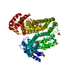
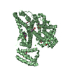
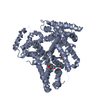
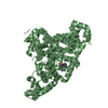
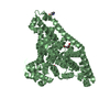
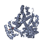
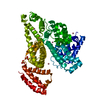
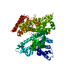
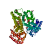
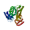
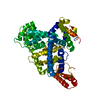
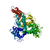
| Title | Crystal Structure of Ovine Serum Albumin | ||||||
 Components Components | Serum albumin | ||||||
 Keywords Keywords | TRANSPORT PROTEIN / Helical structure / transport / Fatty acids / metabolites | ||||||
| Function / homology |  Function and homology informationenterobactin binding / lipid binding / extracellular space / metal ion binding Function and homology informationenterobactin binding / lipid binding / extracellular space / metal ion bindingSimilarity search - Function | ||||||
| Biological species |  Ovis aries (sheep) Ovis aries (sheep) | ||||||
| Method | X-RAY DIFFRACTION / SYNCHROTRON / MOLECULAR REPLACEMENT / Resolution: 2.3 Å | ||||||
 Authors Authors | Bujacz, A. / Talaj, J.A. / Pietrzyk, A.J. / Bujacz, G. | ||||||
 Citation Citation | Journal: To be Published Title: Crystal structure of ovine serum albumin and its complex with 3,5-diiodosalicylic acid Authors: Bujacz, A. / Talaj, J.A. / Pietrzyk, A.J. | ||||||
| History |
|
- Structure visualization
| Structure viewer | Molecule: MolmilJmol/JSmol |
|---|
- Downloads & links
-Download
| PDBx/mmCIF format | 4luf.cif.gz | 248.8 KB | Display | PDBx/mmCIF format |
|---|---|---|---|---|
| PDB format | pdb4luf.ent.gz | 202 KB | Display | PDB format |
| PDBx/mmJSON format | 4luf.json.gz | Tree view | PDBx/mmJSON format | |
| Others |  Other downloads Other downloads |
-Validation report
| Arichive directory | https://data.pdbj.org/pub/pdb/validation_reports/lu/4lufftp://data.pdbj.org/pub/pdb/validation_reports/lu/4luf | HTTPS FTP |
|---|
-Related structure data
| Related structure data | 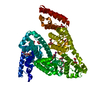 4luhC  4f5sS S: Starting model for refinement C: citing same article ( |
|---|---|
| Similar structure data |


-Links
 PDBj
PDBj


- Assembly
| Deposited unit | 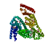
| |||||||||
|---|---|---|---|---|---|---|---|---|---|---|
| 1 |
| |||||||||
| Unit cell |
| |||||||||
| Components on special symmetry positions |
|
-Components
-Protein , 1 types, 1 molecules A
| #1: Protein | Mass: 66427.820 Da / Num. of mol.: 1 / Fragment: Mature form of ovine serum albumin / Source method: isolated from a natural source / Source: (natural) Ovis aries (sheep) / References: UniProt: P14639 |
|---|
-Non-polymers , 7 types, 202 molecules 


| #2: Chemical |  Mass: 250.332 Da / Num. of mol.: 3 / Source method: obtained synthetically / Formula: C12H26O5 Mass: 250.332 Da / Num. of mol.: 3 / Source method: obtained synthetically / Formula: C12H26O5#3: Chemical | ChemComp-LMR / ( | Malic acid Mass: 134.087 Da / Num. of mol.: 1 / Source method: obtained synthetically / Formula: C4H6O5 Mass: 134.087 Da / Num. of mol.: 1 / Source method: obtained synthetically / Formula: C4H6O5#4: Chemical | ChemComp-ACT / | Acetate Mass: 59.044 Da / Num. of mol.: 1 / Source method: obtained synthetically / Formula: C2H3O2 Mass: 59.044 Da / Num. of mol.: 1 / Source method: obtained synthetically / Formula: C2H3O2#5: Chemical | Malonic acid Mass: 102.046 Da / Num. of mol.: 2 / Source method: obtained synthetically / Formula: C3H2O4 Mass: 102.046 Da / Num. of mol.: 2 / Source method: obtained synthetically / Formula: C3H2O4#6: Chemical | Succinic acid Mass: 118.088 Da / Num. of mol.: 3 / Source method: obtained synthetically / Formula: C4H6O4 Mass: 118.088 Da / Num. of mol.: 3 / Source method: obtained synthetically / Formula: C4H6O4#7: Chemical | ChemComp-FMT / | Formic acid Mass: 46.025 Da / Num. of mol.: 1 / Source method: obtained synthetically / Formula: CH2O2 Mass: 46.025 Da / Num. of mol.: 1 / Source method: obtained synthetically / Formula: CH2O2#8: Water | ChemComp-HOH / | WaterMass: 18.015 Da / Num. of mol.: 191 / Source method: isolated from a natural source / Formula: H2O |
|---|
-Experimental details
-Experiment
| Experiment | Method: X-RAY DIFFRACTION / Number of used crystals: 1 |
|---|
- Sample preparation
| Crystal | Density Matthews: 3.68 Å3/Da / Density % sol: 66.57 % |
|---|---|
| Crystal grow | Temperature: 293 K / pH: 7 Details: Tacsimate 70%, 4% PPG 400, pH 7.0, VAPOR DIFFUSION, HANGING DROP, temperature 293K |
-Data collection
| Diffraction | Mean temperature: 100 K |
|---|---|
| Diffraction source | Source: SYNCHROTRON / Site: BESSY  / Beamline: 14.2 / Wavelength: 0.9184 / Beamline: 14.2 / Wavelength: 0.9184 |
| Detector | Type: MARMOSAIC 225 mm CCD / Detector: CCD / Date: Apr 14, 2013 / Details: MIRRORS |
| Radiation | Monochromator: DOUBLE CRYSTAL MONOCHROMATOR SI / Protocol: SINGLE WAVELENGTH / Monochromatic (M) / Laue (L): M / Scattering type: x-ray |
| Radiation wavelength | Wavelength: 0.9184 Å / Relative weight: 1 |
| Reflection | Resolution: 2.3→50 Å / Num. obs: 43652 / % possible obs: 99.4 % / Observed criterion σ(I): -3 / Redundancy: 3.69 % / Biso Wilson estimate: 46.1 Å2 / Rmerge(I) obs: 0.096 / Net I/σ(I): 12.11 |
| Reflection shell | Resolution: 2.3→2.4 Å / Redundancy: 3.73 % / Rmerge(I) obs: 0.902 / Mean I/σ(I) obs: 2 / % possible all: 99.8 |
- Processing
| Software |
| ||||||||||||||||||||||||||||||||||||||||||||||||||||||||||||||||||||||||||||||||||||||||||||||||||||||||||||||||||||||||||||||||||||||||||||||||||||||||||||||||||||||||||||||||||||||
|---|---|---|---|---|---|---|---|---|---|---|---|---|---|---|---|---|---|---|---|---|---|---|---|---|---|---|---|---|---|---|---|---|---|---|---|---|---|---|---|---|---|---|---|---|---|---|---|---|---|---|---|---|---|---|---|---|---|---|---|---|---|---|---|---|---|---|---|---|---|---|---|---|---|---|---|---|---|---|---|---|---|---|---|---|---|---|---|---|---|---|---|---|---|---|---|---|---|---|---|---|---|---|---|---|---|---|---|---|---|---|---|---|---|---|---|---|---|---|---|---|---|---|---|---|---|---|---|---|---|---|---|---|---|---|---|---|---|---|---|---|---|---|---|---|---|---|---|---|---|---|---|---|---|---|---|---|---|---|---|---|---|---|---|---|---|---|---|---|---|---|---|---|---|---|---|---|---|---|---|---|---|---|---|
| Refinement | Method to determine structure: MOLECULAR REPLACEMENT Starting model: PDB ENTRY 4F5S Resolution: 2.3→47.19 Å / Cor.coef. Fo:Fc: 0.961 / Cor.coef. Fo:Fc free: 0.927 / SU B: 10.471 / SU ML: 0.119 / Isotropic thermal model: Isotropic / Cross valid method: THROUGHOUT / σ(F): 0 / ESU R: 0.192 / ESU R Free: 0.169 / Stereochemistry target values: MAXIMUM LIKELIHOOD
| ||||||||||||||||||||||||||||||||||||||||||||||||||||||||||||||||||||||||||||||||||||||||||||||||||||||||||||||||||||||||||||||||||||||||||||||||||||||||||||||||||||||||||||||||||||||
| Solvent computation | Ion probe radii: 0.8 Å / Shrinkage radii: 0.8 Å / VDW probe radii: 1.2 Å / Solvent model: BABINET MODEL WITH MASK | ||||||||||||||||||||||||||||||||||||||||||||||||||||||||||||||||||||||||||||||||||||||||||||||||||||||||||||||||||||||||||||||||||||||||||||||||||||||||||||||||||||||||||||||||||||||
| Displacement parameters | Biso mean: 47.65 Å2
| ||||||||||||||||||||||||||||||||||||||||||||||||||||||||||||||||||||||||||||||||||||||||||||||||||||||||||||||||||||||||||||||||||||||||||||||||||||||||||||||||||||||||||||||||||||||
| Refinement step | Cycle: LAST / Resolution: 2.3→47.19 Å
| ||||||||||||||||||||||||||||||||||||||||||||||||||||||||||||||||||||||||||||||||||||||||||||||||||||||||||||||||||||||||||||||||||||||||||||||||||||||||||||||||||||||||||||||||||||||
| Refine LS restraints |
|