 Movie
Movie Controller

[English] 日本語
 Yorodumi
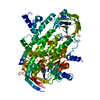
- PDB-6es9: Methylsuccinyl-CoA dehydrogenase of Paracoccus denitrificans with... -
+ Open data

- Basic information
| Entry | Database: PDB / ID: 6es9 | ||||||
|---|---|---|---|---|---|---|---|
| Title | Methylsuccinyl-CoA dehydrogenase of Paracoccus denitrificans with bound flavin adenine dinucleotide | ||||||
 Components Components | Acyl-CoA dehydrogenase | ||||||
 Keywords Keywords | FLAVOPROTEIN / methylsuccinyl-CoA / acyl-CoA dehydrogenase / flavin adenine dinucleotide / ethylmalonyl-CoA pathway | ||||||
| Function / homology |  Function and homology informationOxidoreductases; Acting on the CH-CH group of donors; With a flavin as acceptor / acyl-CoA dehydrogenase activity / flavin adenine dinucleotide binding Function and homology informationOxidoreductases; Acting on the CH-CH group of donors; With a flavin as acceptor / acyl-CoA dehydrogenase activity / flavin adenine dinucleotide bindingSimilarity search - Function | ||||||
| Biological species |  Paracoccus denitrificans (bacteria) Paracoccus denitrificans (bacteria) | ||||||
| Method | X-RAY DIFFRACTION / SYNCHROTRON / MOLECULAR REPLACEMENT / molecular replacement / Resolution: 1.37 Å | ||||||
 Authors Authors | Zarzycki, J. / Schwander, T. / Erb, T.J. | ||||||
 Citation Citation | Journal: J. Biol. Chem. / Year: 2018 Title: Structural basis for substrate specificity of methylsuccinyl-CoA dehydrogenase, an unusual member of the acyl-CoA dehydrogenase family. Authors: Schwander, T. / McLean, R. / Zarzycki, J. / Erb, T.J. | ||||||
| History |
|
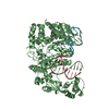
- Structure visualization
| Structure viewer | Molecule: MolmilJmol/JSmol |
|---|
- Downloads & links
-Download
| PDBx/mmCIF format | 6es9.cif.gz | 486.6 KB | Display | PDBx/mmCIF format |
|---|---|---|---|---|
| PDB format | pdb6es9.ent.gz | 393.1 KB | Display | PDB format |
| PDBx/mmJSON format | 6es9.json.gz | Tree view | PDBx/mmJSON format | |
| Others |  Other downloads Other downloads |
-Validation report
| Arichive directory | https://data.pdbj.org/pub/pdb/validation_reports/es/6es9ftp://data.pdbj.org/pub/pdb/validation_reports/es/6es9 | HTTPS FTP |
|---|
-Related structure data
| Related structure data | 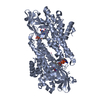 4n5fS S: Starting model for refinement |
|---|---|
| Similar structure data |

-Links
 PDBj
PDBj


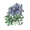
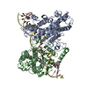
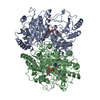
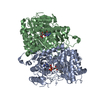
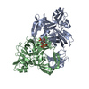
- Assembly
| Deposited unit | 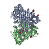
| ||||||||
|---|---|---|---|---|---|---|---|---|---|
| 1 |
| ||||||||
| Unit cell |
|
-Components
| #1: Protein | Mass: 59985.164 Da / Num. of mol.: 2 Source method: isolated from a genetically manipulated source Source: (gene. exp.) Paracoccus denitrificans (strain Pd 1222) (bacteria)Strain: Pd 1222 / Gene: Pden_2840 / Plasmid: pTE849 / Production host: Escherichia coli BL21(DE3) (bacteria)References: UniProt: A1B5Y0, Oxidoreductases; Acting on the CH-CH group of donors; With a flavin as acceptor#2: Chemical | ChemComp-SO4 / Sulfate  Mass: 96.063 Da / Num. of mol.: 4 / Source method: obtained synthetically / Formula: SO4 Mass: 96.063 Da / Num. of mol.: 4 / Source method: obtained synthetically / Formula: SO4#3: Chemical | Coenzyme A  Mass: 767.534 Da / Num. of mol.: 2 / Source method: obtained synthetically / Formula: C21H36N7O16P3S Mass: 767.534 Da / Num. of mol.: 2 / Source method: obtained synthetically / Formula: C21H36N7O16P3S#4: Chemical | Flavin adenine dinucleotide  Mass: 785.550 Da / Num. of mol.: 2 / Source method: obtained synthetically / Formula: C27H33N9O15P2 / Comment: FAD*YM Mass: 785.550 Da / Num. of mol.: 2 / Source method: obtained synthetically / Formula: C27H33N9O15P2 / Comment: FAD*YM#5: Water | ChemComp-HOH / | Water Mass: 18.015 Da / Num. of mol.: 1389 / Source method: isolated from a natural source / Formula: H2O Mass: 18.015 Da / Num. of mol.: 1389 / Source method: isolated from a natural source / Formula: H2O |
|---|
-Experimental details
-Experiment
| Experiment | Method: X-RAY DIFFRACTION / Number of used crystals: 1 |
|---|
- Sample preparation
| Crystal | Density Matthews: 2.49 Å3/Da / Density % sol: 50.51 % |
|---|---|
| Crystal grow | Temperature: 289 K / Method: vapor diffusion, sitting drop / pH: 8 Details: 1 part protein solution (20.0 mg/mL in 20 mM Tris-HCl, pH 7.9, 200 mM sodium chloride, 1.5 mM FAD, 3 mM mesaconyl-CoA) + 1 part buffer (0.1 M Tris-HCl, pH 8.0, 30% w/v PEG5000 MME, 200 mM lithium sulfate) |
-Data collection
| Diffraction | Mean temperature: 100 K | ||||||||||||||||||||||||||||||
|---|---|---|---|---|---|---|---|---|---|---|---|---|---|---|---|---|---|---|---|---|---|---|---|---|---|---|---|---|---|---|---|
| Diffraction source | Source: SYNCHROTRON / Site: ESRF  / Beamline: ID29 / Wavelength: 0.979026 Å / Beamline: ID29 / Wavelength: 0.979026 Å | ||||||||||||||||||||||||||||||
| Detector | Type: DECTRIS PILATUS 6M / Detector: PIXEL / Date: Jun 26, 2017 | ||||||||||||||||||||||||||||||
| Radiation | Protocol: SINGLE WAVELENGTH / Monochromatic (M) / Laue (L): M / Scattering type: x-ray | ||||||||||||||||||||||||||||||
| Radiation wavelength | Wavelength: 0.979026 Å / Relative weight: 1 | ||||||||||||||||||||||||||||||
| Reflection | Resolution: 1.37→17.573 Å / Num. obs: 249985 / % possible obs: 100 % / Redundancy: 7.4 % / Biso Wilson estimate: 15.84 Å2 / CC1/2: 0.999 / Rpim(I) all: 0.026 / Rrim(I) all: 0.07 / Rsym value: 0.065 / Net I/av σ(I): 7.7 / Net I/σ(I): 15.2 | ||||||||||||||||||||||||||||||
| Reflection shell | Diffraction-ID: 1
|
-Phasing
| Phasing | Method: molecular replacement |
|---|
- Processing
| Software |
| ||||||||||||||||||||||||||||||||||||||||||||||||||||||||||||||||||||||||||||||||||||||||||
|---|---|---|---|---|---|---|---|---|---|---|---|---|---|---|---|---|---|---|---|---|---|---|---|---|---|---|---|---|---|---|---|---|---|---|---|---|---|---|---|---|---|---|---|---|---|---|---|---|---|---|---|---|---|---|---|---|---|---|---|---|---|---|---|---|---|---|---|---|---|---|---|---|---|---|---|---|---|---|---|---|---|---|---|---|---|---|---|---|---|---|---|
| Refinement | Method to determine structure: MOLECULAR REPLACEMENT Starting model: 4N5F Resolution: 1.37→17.573 Å / SU ML: 0.11 / Cross valid method: FREE R-VALUE / σ(F): 1.33 / Phase error: 19.44
| ||||||||||||||||||||||||||||||||||||||||||||||||||||||||||||||||||||||||||||||||||||||||||
| Solvent computation | Shrinkage radii: 0.9 Å / VDW probe radii: 1.11 Å | ||||||||||||||||||||||||||||||||||||||||||||||||||||||||||||||||||||||||||||||||||||||||||
| Displacement parameters | Biso max: 71.5 Å2 / Biso mean: 22.0831 Å2 / Biso min: 8.82 Å2 | ||||||||||||||||||||||||||||||||||||||||||||||||||||||||||||||||||||||||||||||||||||||||||
| Refinement step | Cycle: final / Resolution: 1.37→17.573 Å
| ||||||||||||||||||||||||||||||||||||||||||||||||||||||||||||||||||||||||||||||||||||||||||
| Refine LS restraints |
| ||||||||||||||||||||||||||||||||||||||||||||||||||||||||||||||||||||||||||||||||||||||||||
| LS refinement shell | Refine-ID: X-RAY DIFFRACTION / Rfactor Rfree error: 0 / Total num. of bins used: 14 / % reflection obs: 100 %
| ||||||||||||||||||||||||||||||||||||||||||||||||||||||||||||||||||||||||||||||||||||||||||
| Refinement TLS params. | Method: refined / Origin x: 9.0776 Å / Origin y: 0.3976 Å / Origin z: -14.9819 Å
| ||||||||||||||||||||||||||||||||||||||||||||||||||||||||||||||||||||||||||||||||||||||||||
| Refinement TLS group |
|
