- PDB-1o75: Tp47, the 47-Kilodalton Lipoprotein of Treponema pallidum -
+
Open data
ID or keywords:
Loading...
-
Basic information
Entry
Database: PDB / ID: 1o75
Title
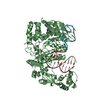
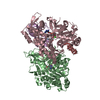
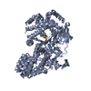
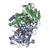
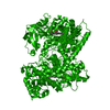
Tp47, the 47-Kilodalton Lipoprotein of Treponema pallidum
Components
47 KDA MEMBRANE ANTIGEN
Keywords
LIPOPROTEINULLNTIGEN / PENICILLIN-BINDING PROTEIN / INTEGRAL MEMBRANE LIPOPROTEIN / IMMUNOGEN / FOUR-DOMAIN PROTEIN / ANTIGEN / LIPOPROTEIN
Function / homology
Function and homology information
Hydrolases; Acting on peptide bonds (peptidases) / peptidase activity / plasma membrane Similarity search - Function
Penicillin-binding protein Tp47, domain B / Penicillin-binding protein Tp47, domain D / Penicillin-binding protein Tp47, domain C / Penicillin-binding protein Tp47, domain A / Penicillin-binding protein Tp47, domain C / Penicillin-binding protein Tp47, domain D / Penicillin-binding protein Tp47 domain A / Tp47, N-terminal domain superfamily / Tp47 lipoprotein, middle and C-terminal domain / Tp47, domain C superfamily ...Penicillin-binding protein Tp47, domain B / Penicillin-binding protein Tp47, domain D / Penicillin-binding protein Tp47, domain C / Penicillin-binding protein Tp47, domain A / Penicillin-binding protein Tp47, domain C / Penicillin-binding protein Tp47, domain D / Penicillin-binding protein Tp47 domain A / Tp47, N-terminal domain superfamily / Tp47 lipoprotein, middle and C-terminal domain / Tp47, domain C superfamily / Penicillin-binding protein Tp47 domain C / Penicillin-binding protein Tp47 domain a / Dna Ligase; domain 1 / SH3 type barrels. / Prokaryotic membrane lipoprotein lipid attachment site profile. / Roll / Immunoglobulin-like / Sandwich / 2-Layer Sandwich / Mainly Beta / Alpha Beta Similarity search - Domain/homology
#1: Journal: Proc.Natl.Acad.Sci. USA / Year: 1994 Title: The 47-kDa Major Lipoprotein Immunogen of Treponema Pallidum is a Penicillin-Binding Protein with Carboxypeptidase Activity Authors: Weigel, L.M. / Radolf, J.D. / Norgard, M.V.
SHEET THE SHEET STRUCTURE OF THIS MOLECULE IS BIFURCATED. IN ORDER TO REPRESENT THIS FEATURE IN ... SHEET THE SHEET STRUCTURE OF THIS MOLECULE IS BIFURCATED. IN ORDER TO REPRESENT THIS FEATURE IN THE SHEET RECORDS BELOW, TWO SHEETS ARE DEFINED.
THE OLIGOMERIZATION STATE OF THE PROTEIN IN SOLUTIONIS MONOMERIC, AS DETERMINED VIA ANALYTICALULTRACENTRIFUGATION. THE BIOLOGICALLY SIGNIFICANTOLIGOMERIZATION STATE OF THE MEMBRANCE BOUNDLIPOPROTIEN IS UNKNOWN
-
Components
#1: Protein
47KDAMEMBRANEANTIGEN
Mass: 45664.566 Da / Num. of mol.: 2 / Mutation: YES Source method: isolated from a genetically manipulated source Source: (gene. exp.) TREPONEMA PALLIDUM (bacteria) / Plasmid: PASK-IBA7 / Production host: Escherichia coli DH5[alpha] (bacteria) / References: UniProt: P29723
Mass: 18.015 Da / Num. of mol.: 619 / Source method: isolated from a natural source / Formula: H2O
Compound details
THIS MEMBRANE LIPOPROTEIN IS A PATHOGEN-SPECIFIC MEMBRANE IMMUNOGEN FOUND ONLY IN TREPONEMES. MAY ...THIS MEMBRANE LIPOPROTEIN IS A PATHOGEN-SPECIFIC MEMBRANE IMMUNOGEN FOUND ONLY IN TREPONEMES. MAY PLAY AN IMPORTANT ROLE IN THE CELL ENVELOPE OF VIRULENT TREPONEMA. ENGINEERED MUTATION HIS 24 SER AND HIS 28 SER, CHAINS A, B
Sequence details
THE FIRST 20 RESIDUES OF THIS SEQUENCE ENCODES FOR THE LIPIDATED MEMBRANE ANCHOR. THE POST- ...THE FIRST 20 RESIDUES OF THIS SEQUENCE ENCODES FOR THE LIPIDATED MEMBRANE ANCHOR. THE POST-TRANSLATIONALLY MODIFIED N-TERMINAL CYSTEINE OF NATIVE TP47 WAS DESIGNATED AS AMINO ACID #1. ONLY RESIDUES 2-415 WERE EXPRESSED.
-
Experimental details
-
Experiment
Experiment
Method: X-RAY DIFFRACTION / Number of used crystals: 1
-
Sample preparation
Crystal
Density Matthews: 3.97 Å3/Da / Density % sol: 45 %
Crystal grow
pH: 5.6 Details: 32% PEG 4000, 100 MM SODIUM CITRATE PH 5.6, 200 MM AMMONIUM ACETATE,3% (W/V) DEXTRAN SULFATE 8000
In the structure databanks used in Yorodumi, some data are registered as the other names, "COVID-19 virus" and "2019-nCoV". Here are the details of the virus and the list of structure data.
Jan 31, 2019. EMDB accession codes are about to change! (news from PDBe EMDB page)
EMDB accession codes are about to change! (news from PDBe EMDB page)
The allocation of 4 digits for EMDB accession codes will soon come to an end. Whilst these codes will remain in use, new EMDB accession codes will include an additional digit and will expand incrementally as the available range of codes is exhausted. The current 4-digit format prefixed with “EMD-” (i.e. EMD-XXXX) will advance to a 5-digit format (i.e. EMD-XXXXX), and so on. It is currently estimated that the 4-digit codes will be depleted around Spring 2019, at which point the 5-digit format will come into force.
The EM Navigator/Yorodumi systems omit the EMD- prefix.
Related info.:Q: What is EMD? / ID/Accession-code notation in Yorodumi/EM Navigator
Yorodumi is a browser for structure data from EMDB, PDB, SASBDB, etc.
This page is also the successor to EM Navigator detail page, and also detail information page/front-end page for Omokage search.
The word "yorodu" (or yorozu) is an old Japanese word meaning "ten thousand". "mi" (miru) is to see.
Related info.:EMDB / PDB / SASBDB / Comparison of 3 databanks / Yorodumi Search / Aug 31, 2016. New EM Navigator & Yorodumi / Yorodumi Papers / Jmol/JSmol / Function and homology information / Changes in new EM Navigator and Yorodumi
 Movie
Movie Controller


 Open data
 Basic information
 Components
 Keywords
 PENICILLIN-BINDING PROTEIN / INTEGRAL MEMBRANE LIPOPROTEIN /
  Function and homology information
 Authors
 Citation
 Structure visualization
 Downloads & links
 Other downloads
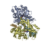


 PDBj
PDBj


 Assembly
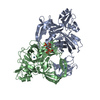

 Mass: 131.293 Da / Num. of mol.: 5 / Source method: obtained synthetically / Formula: Xe
 Mass: 18.015 Da / Num. of mol.: 619 / Source method: isolated from a natural source / Formula: H2O
 Sample preparation
 / Beamline: 19-ID / Wavelength: 0.97938
  Processing