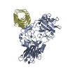
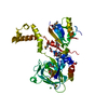
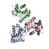
Entry Database : PDB / ID : 6es1Title Crystal structure of the binding domain from botulinum neurotoxin A2 bound to extracellular domain of human receptor SV2C Botulinum neurotoxin type A Synaptic vesicle glycoprotein 2C Keywords / / / / / Function / homology Function Domain/homology Component
/ / / / / / / / / / / / / / / / / / / / / / / / / / / / / / / / / / / / / / / / / / / / / / / / / / / / / / / / / / / / / / / / / / / / / / / Biological species Clostridium botulinum (bacteria)Homo sapiens (human)Method / / / Resolution : 2 Å Authors Gustafsson, R. / Masuyer, G. / Stenmark, P. Funding support Organization Grant number Country Swedish Research Council 2014-5667 Wenner-Gren Foundation Swedish Cancer Society National Institutes of Health 1R01NS080833
Journal : Toxins (Basel) / Year : 2018Title : Crystal Structure of Botulinum Neurotoxin A2 in Complex with the Human Protein Receptor SV2C Reveals Plasticity in Receptor Binding.Authors : Gustafsson, R. / Zhang, S. / Masuyer, G. / Dong, M. / Stenmark, P. History Deposition Oct 19, 2017 Deposition site / Processing site Revision 1.0 Apr 25, 2018 Provider / Type Revision 1.1 Jan 17, 2024 Group / Database references / Refinement descriptionCategory chem_comp_atom / chem_comp_bond ... chem_comp_atom / chem_comp_bond / database_2 / pdbx_initial_refinement_model Item / _database_2.pdbx_database_accession
Show all Show less
 Movie
Movie Controller

 Yorodumi
 Open data
 Basic information
 Components
 Keywords
 PROTEIN BINDING / Receptor /
  Function and homology information


 Authors
 Sweden,
  United States, 4items
  Citation
 Structure visualization
 Downloads & links
 Other downloads
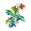


 PDBj
PDBj


 Assembly

 Mass: 59.044 Da / Num. of mol.: 1 / Source method: obtained synthetically / Formula: C2H3O2
 Mass: 18.015 Da / Num. of mol.: 359 / Source method: isolated from a natural source / Formula: H2O
 Sample preparation
 / Beamline: 14.1 / Wavelength: 0.919908 Å
 Processing