 ムービー
ムービー コントローラー


+ データを開く

- 基本情報
基本情報
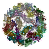
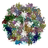
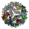
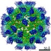
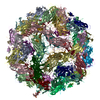
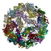
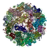
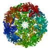
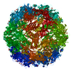
| 登録情報 | データベース: PDB / ID: 6e2z | |||||||||
|---|---|---|---|---|---|---|---|---|---|---|
| タイトル | Mechanism of cellular recognition by PCV2 | |||||||||
 要素 要素 | Capsid protein of PCV2 カプシド カプシド | |||||||||
 キーワード キーワード | VIRUS LIKE PARTICLE (ウイルス様粒子) / viral jelly-roll | |||||||||
| 機能・相同性 | Circovirus capsid protein / Circovirus capsid superfamily / Circovirus capsid protein / viral capsid assembly / T=1 icosahedral viral capsid / symbiont entry into host cell / virion attachment to host cell / Cap 機能・相同性情報 機能・相同性情報 | |||||||||
| 生物種 |   Porcine circovirus 2 (ウイルス) Porcine circovirus 2 (ウイルス) | |||||||||
| 手法 | 電子顕微鏡法 / 単粒子再構成法 / クライオ電子顕微鏡法 / 解像度: 3.4 Å | |||||||||
| Model details | Cryo-EM image reconstruction at 3.2Angstrom resolution | |||||||||
 データ登録者 データ登録者 | Khayat, R. / Dhindwal, S. | |||||||||
| 資金援助 |  米国, 1件 米国, 1件
| |||||||||
 引用 引用 | ジャーナル: J Virol / 年: 2019 タイトル: Porcine Circovirus 2 Uses a Multitude of Weak Binding Sites To Interact with Heparan Sulfate, and the Interactions Do Not Follow the Symmetry of the Capsid. 著者: Sonali Dhindwal / Bryant Avila / Shanshan Feng / Reza Khayat / 要旨: Porcine circovirus 2 (PCV2) is the smallest pathogenic virus capable of autonomous replication within its host. Infections result in immunosuppression and subsequent death of the host and are ...Porcine circovirus 2 (PCV2) is the smallest pathogenic virus capable of autonomous replication within its host. Infections result in immunosuppression and subsequent death of the host and are initiated via the attachment of the PCV2 icosahedral capsid to heparan sulfate (HS) and chondroitin sulfate B (CSB) glycosaminoglycans on the cell surface. However, the underlying mechanism of structural recognition remains to be explored. Using heparin, a routinely used analog of heparan sulfate, we demonstrate that increasing lengths of heparin exhibit a greater affinity toward PCV2. Our competition assays indicate that dextran sulfate (8 kDa) has a higher affinity for PCV2 than heparin (12 kDa), chondroitin sulfate B (41 kDa), hyaluronic acid (1.6 MDa), and dextran (6 kDa). This suggests that polymers high in sulfate content are capable of competing with the PCV2-heparan sulfate interaction and, thus, have the potential to inhibit PCV2 infection. Finally, we visualized the interaction between heparin and the PCV2 capsid using cryo-electron microscopy single-particle analysis, symmetry expansion, and focused classification. The image reconstructions provide the first example of an asymmetric distribution of heparin on the surface of an icosahedral virus capsid. We demonstrate that each of the 60 capsid subunits that generate the T1 capsid can bind heparin via one of five binding sites. However, not all of the binding sites were occupied by heparin, and only one-third to two-thirds of the binding sites were occupied. The binding sites are defined by arginine, lysine, and polar amino acids. Mutating the arginine, lysine, and polar amino acids to alanine diminished the binding capacity of PCV2 to heparin. It has been demonstrated that porcine circovirus 2 (PCV2) attaches to cells via heparan sulfate (HS) and chondroitin sulfate B (CSB) glycosaminoglycans; however, the underlying structural mechanism describing the HS/CSB recognition by PCV2 remains to be explored. We used cryo-electron microscopy with single-particle analysis, symmetry expansion, and focused classification to visualize the interaction between the PCV2 capsid and heparin, an analog of heparan sulfate, to better than 3.6-Å resolution. We observed that the interaction between PCV2 and heparin does not adhere to the icosahedral symmetry of the capsid. To the best of our knowledge, this is the first example where the interaction between heparin and an icosahedral capsid does not follow the symmetry elements of the capsid. Our findings also suggest that anionic polymers, such as dextran sulfate, may act to inhibit PCV2 infection. | |||||||||
| 履歴 |
|
- 構造の表示
構造の表示
| ムービー |
ムービービューア |
|---|---|
| 構造ビューア | 分子: MolmilJmol/JSmol |
- ダウンロードとリンク
-ダウンロード
| PDBx/mmCIF形式 | 6e2z.cif.gz | 3.6 MB | 表示 | PDBx/mmCIF形式 |
|---|---|---|---|---|
| PDB形式 | pdb6e2z.ent.gz | 表示 | PDB形式 | |
| PDBx/mmJSON形式 | 6e2z.json.gz | ツリー表示 | PDBx/mmJSON形式 | |
| その他 |  その他のダウンロード その他のダウンロード |
-検証レポート
| アーカイブディレクトリ | https://data.pdbj.org/pub/pdb/validation_reports/e2/6e2zftp://data.pdbj.org/pub/pdb/validation_reports/e2/6e2z | HTTPS FTP |
|---|
-関連構造データ
| 関連構造データ | 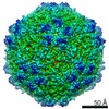 8971MC 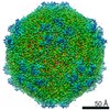 8939C 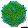 8969C 8970C 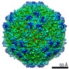 8972C 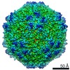 8973C 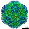 8974C 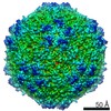 8975C 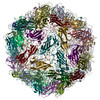 6dzuC 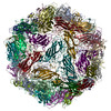 6e2rC 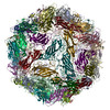 6e2xC  6e30C 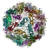 6e32C  6e34C 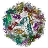 6e39C C: 同じ文献を引用 ( M: このデータのモデリングに利用したマップデータ |
|---|---|
| 類似構造データ |

-リンク
 PDBj
PDBj
- 集合体
集合体
| 登録構造単位 | 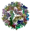
|
|---|---|
| 1 |
|
-要素
| #1: タンパク質 | カプシド 分子量: 27899.795 Da / 分子数: 60 / 由来タイプ: 組換発現 / 由来: (組換発現) Porcine circovirus 2 (ウイルス) / 遺伝子: cap, Cap, cp, ORF2, PCV2_gp3 / プラスミド: pFastBac1 / 発現宿主:  Trichoplusia ni (イラクサキンウワバ) / 株 (発現宿主): High Five / 参照: UniProt: Q805N7 Trichoplusia ni (イラクサキンウワバ) / 株 (発現宿主): High Five / 参照: UniProt: Q805N7#2: 多糖 | 2-deoxy-6-O-sulfo-2-(sulfoamino)-alpha-D-glucopyranose-(1-4)-2-O-sulfo-alpha-L-idopyranuronic acid- ...2-deoxy-6-O-sulfo-2-(sulfoamino)-alpha-D-glucopyranose-(1-4)-2-O-sulfo-alpha-L-idopyranuronic acid-(1-4)-2-deoxy-6-O-sulfo-2-(sulfoamino)-alpha-D-glucopyranose | オリゴ糖 / 分子量: 916.769 Da / 分子数: 1 / 由来タイプ: 組換発現 |
|---|
-実験情報
-実験
| 実験 | 手法: 電子顕微鏡法 |
|---|---|
| EM実験 | 試料の集合状態: PARTICLE / 3次元再構成法: 単粒子再構成法 |
- 試料調製
試料調製
| 構成要素 |
| ||||||||||||||||||||||||||||
|---|---|---|---|---|---|---|---|---|---|---|---|---|---|---|---|---|---|---|---|---|---|---|---|---|---|---|---|---|---|
| 分子量 |
| ||||||||||||||||||||||||||||
| 由来(天然) |
| ||||||||||||||||||||||||||||
| 由来(組換発現) |
| ||||||||||||||||||||||||||||
| ウイルスについての詳細 |
| ||||||||||||||||||||||||||||
| 天然宿主 |
| ||||||||||||||||||||||||||||
| ウイルス殻 |
| ||||||||||||||||||||||||||||
| 緩衝液 | pH: 7 | ||||||||||||||||||||||||||||
| 緩衝液成分 |
| ||||||||||||||||||||||||||||
| 試料 | 濃度: 0.718 mg/ml / 包埋: NO / シャドウイング: NO / 染色: NO / 凍結: YES / 詳細: This sample was monodisperse | ||||||||||||||||||||||||||||
| 試料支持 | 詳細: unspecified | ||||||||||||||||||||||||||||
| 急速凍結 | 装置: FEI VITROBOT MARK IV / 凍結剤: ETHANE / 湿度: 100 % / 凍結前の試料温度: 4 K |

- 電子顕微鏡撮影
| 顕微鏡 | モデル: FEI TITAN |
|---|---|
| 電子銃 | 電子線源: FIELD EMISSION GUN / 加速電圧: 300 kV / 照射モード: FLOOD BEAM |
| 電子レンズ | モード: BRIGHT FIELDBright-field microscopy / Cs: 0.01 mm / C2レンズ絞り径: 100 µm / アライメント法: COMA FREE |
| 試料ホルダ | 凍結剤: NITROGEN |
| 撮影 | 平均露光時間: 5 sec. / 電子線照射量: 32 e/Å2 / 検出モード: COUNTING フィルム・検出器のモデル: GATAN K2 SUMMIT (4k x 4k) 撮影したグリッド数: 1 / 実像数: 3725 |
| 画像スキャン | 動画フレーム数/画像: 25 / 利用したフレーム数/画像: 2-25 |
- 解析
解析
| EMソフトウェア |
| |||||||||||||||||||||||||||||||||||||||||||||
|---|---|---|---|---|---|---|---|---|---|---|---|---|---|---|---|---|---|---|---|---|---|---|---|---|---|---|---|---|---|---|---|---|---|---|---|---|---|---|---|---|---|---|---|---|---|---|
| CTF補正 | 詳細: Per particle estimation / タイプ: PHASE FLIPPING AND AMPLITUDE CORRECTION | |||||||||||||||||||||||||||||||||||||||||||||
| 粒子像の選択 | 選択した粒子像数: 166369 | |||||||||||||||||||||||||||||||||||||||||||||
| 対称性 | 点対称性: C1 (非対称) | |||||||||||||||||||||||||||||||||||||||||||||
| 3次元再構成 | 解像度: 3.4 Å / 解像度の算出法: FSC 0.143 CUT-OFF / 粒子像の数: 90206 詳細: Focused classification of symmetry expanded data set. Thus, the number of particles used is equal to the number of subunits present. 対称性のタイプ: POINT | |||||||||||||||||||||||||||||||||||||||||||||
| 原子モデル構築 | B value: 35.5 / プロトコル: FLEXIBLE FIT / 空間: REAL / Target criteria: Correlation coefficient / 詳細: Several iterations of refinement |
