 Movie
Movie Controller

[English] 日本語
 Yorodumi
- PDB-6e09: Crystal Structure of Helicobacter pylori TlpA Chemoreceptor Ligan... -
+ Open data

- Basic information
| Entry | Database: PDB / ID: 6000000000 | ||||||
|---|---|---|---|---|---|---|---|
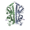
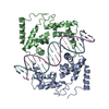
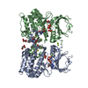
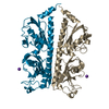
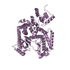
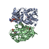
| Title | Crystal Structure of Helicobacter pylori TlpA Chemoreceptor Ligand Binding Domain | ||||||
 Components Components | Methyl-accepting chemotaxis protein TlpA | ||||||
 Keywords Keywords |  SIGNALING PROTEIN / chemoreceptor / helix bundle / PAS/Cache SIGNALING PROTEIN / chemoreceptor / helix bundle / PAS/Cache | ||||||
| Function / homology | Methyl-accepting chemotaxis protein (MCP) signalling domain / Methyl-accepting chemotaxis protein (MCP) signalling domain / Bacterial chemotaxis sensory transducers domain profile. / Methyl-accepting chemotaxis-like domains (chemotaxis sensory transducer). / signal transduction / membrane / metal ion binding / Methyl-accepting chemotaxis protein TlpA Function and homology information Function and homology information | ||||||
| Biological species |  Helicobacter pylori SS1 (bacteria) Helicobacter pylori SS1 (bacteria) | ||||||
| Method | X-RAY DIFFRACTION / SYNCHROTRON / SIRAS / Resolution: 2.3 Å | ||||||
 Authors Authors | Remington, S.J. / Guillemin, K. / Sweeney, E. / Perkins, A. | ||||||
| Funding support |  United States, 1items United States, 1items
| ||||||
 Citation Citation | Journal: Protein Sci. / Year: 2018 Title: Structures of the ligand-binding domain of Helicobacter pylori chemoreceptor TlpA. Authors: Sweeney, E.G. / Perkins, A. / Kallio, K. / James Remington, S. / Guillemin, K. | ||||||
| History |
|
- Structure visualization
| Structure viewer | Molecule: MolmilJmol/JSmol |
|---|
- Downloads & links
-Download
| PDBx/mmCIF format | 6e09.cif.gz | 218.9 KB | Display | PDBx/mmCIF format |
|---|---|---|---|---|
| PDB format | pdb6e09.ent.gz | 182.5 KB | Display | PDB format |
| PDBx/mmJSON format | 6e09.json.gz | Tree view | PDBx/mmJSON format | |
| Others |  Other downloads Other downloads |
-Validation report
| Arichive directory | https://data.pdbj.org/pub/pdb/validation_reports/e0/6e09ftp://data.pdbj.org/pub/pdb/validation_reports/e0/6e09 | HTTPS FTP |
|---|
-Related structure data


-Links
 PDBj
PDBj
- Assembly
| Deposited unit | 
| ||||||||
|---|---|---|---|---|---|---|---|---|---|
| 1 | 
| ||||||||
| 2 | 
| ||||||||
| Unit cell |
|
-Components
| #1: Protein | Mass: 34068.559 Da / Num. of mol.: 4 Source method: isolated from a genetically manipulated source Source: (gene. exp.) Helicobacter pylori SS1 (bacteria) / Gene: tlpA, HPYLSS1_00094 / Production host: Escherichia coli (E. coli) / Strain (production host): JM109 / Variant (production host): DE3 / References: UniProt: A0A1U9IS38#2: Water | ChemComp-HOH / | Water Mass: 18.015 Da / Num. of mol.: 529 / Source method: isolated from a natural source / Formula: H2O Mass: 18.015 Da / Num. of mol.: 529 / Source method: isolated from a natural source / Formula: H2O |
|---|
-Experimental details
-Experiment
| Experiment | Method: X-RAY DIFFRACTION / Number of used crystals: 1 |
|---|
- Sample preparation
| Crystal | Density Matthews: 2.27 Å3/Da / Density % sol: 45.85 % |
|---|---|
| Crystal grow | Temperature: 277 K / Method: vapor diffusion, hanging drop / pH: 7.5 Details: 19% PEG 3350, 0.15M AmNO3, 0.15M NaCl, 25mM HEPES pH 7.5, 2mM DTT |
-Data collection
| Diffraction | Mean temperature: 100 K |
|---|---|
| Diffraction source | Source: SYNCHROTRON / Site: APS / Beamline: 34-ID / Wavelength: 0.97 Å |
| Detector | Type: ADSC QUANTUM 4 / Detector: CCD / Date: Jul 18, 2014 |
| Radiation | Protocol: SINGLE WAVELENGTH / Monochromatic (M) / Laue (L): M / Scattering type: x-ray |
| Radiation wavelength | Wavelength: 0.97 Å / Relative weight: 1 |
| Reflection | Resolution: 2.3→27 Å / Num. obs: 97102 / % possible obs: 99.9 % / Redundancy: 23 % / CC1/2: 0.998 / Rmerge(I) obs: 0.15 / Rpim(I) all: 0.059 / Net I/av σ(I): 13.2 / Net I/σ(I): 13.2 |
| Reflection shell | Resolution: 2.3→2.34 Å / Rmerge(I) obs: 1.75 / Mean I/σ(I) obs: 4.3 / CC1/2: 0.948 / Rpim(I) all: 0.703 / % possible all: 99.6 |
- Processing
| Software |
| ||||||||||||||||||||||||||||||||||||||||||||||||||||||||||||||||||||||||||||||||||||||||||||||||||||||||||||||||||||||||||||||||||||||||||||||||||||||||||||||||||||||||||||||||||||||||||||||||||||
|---|---|---|---|---|---|---|---|---|---|---|---|---|---|---|---|---|---|---|---|---|---|---|---|---|---|---|---|---|---|---|---|---|---|---|---|---|---|---|---|---|---|---|---|---|---|---|---|---|---|---|---|---|---|---|---|---|---|---|---|---|---|---|---|---|---|---|---|---|---|---|---|---|---|---|---|---|---|---|---|---|---|---|---|---|---|---|---|---|---|---|---|---|---|---|---|---|---|---|---|---|---|---|---|---|---|---|---|---|---|---|---|---|---|---|---|---|---|---|---|---|---|---|---|---|---|---|---|---|---|---|---|---|---|---|---|---|---|---|---|---|---|---|---|---|---|---|---|---|---|---|---|---|---|---|---|---|---|---|---|---|---|---|---|---|---|---|---|---|---|---|---|---|---|---|---|---|---|---|---|---|---|---|---|---|---|---|---|---|---|---|---|---|---|---|---|---|---|
| Refinement | Method to determine structure: SIRAS / Resolution: 2.3→26.703 Å / SU ML: 0.28 / Cross valid method: FREE R-VALUE / σ(F): 1.34 / Phase error: 25.4
| ||||||||||||||||||||||||||||||||||||||||||||||||||||||||||||||||||||||||||||||||||||||||||||||||||||||||||||||||||||||||||||||||||||||||||||||||||||||||||||||||||||||||||||||||||||||||||||||||||||
| Solvent computation | Shrinkage radii: 0.9 Å / VDW probe radii: 1.11 Å | ||||||||||||||||||||||||||||||||||||||||||||||||||||||||||||||||||||||||||||||||||||||||||||||||||||||||||||||||||||||||||||||||||||||||||||||||||||||||||||||||||||||||||||||||||||||||||||||||||||
| Refinement step | Cycle: LAST / Resolution: 2.3→26.703 Å
| ||||||||||||||||||||||||||||||||||||||||||||||||||||||||||||||||||||||||||||||||||||||||||||||||||||||||||||||||||||||||||||||||||||||||||||||||||||||||||||||||||||||||||||||||||||||||||||||||||||
| Refine LS restraints |
| ||||||||||||||||||||||||||||||||||||||||||||||||||||||||||||||||||||||||||||||||||||||||||||||||||||||||||||||||||||||||||||||||||||||||||||||||||||||||||||||||||||||||||||||||||||||||||||||||||||
| LS refinement shell |
|
