 Movie
Movie Controller


+ Open data

- Basic information
| Entry | Database: PDB / ID: 6dte | ||||||
|---|---|---|---|---|---|---|---|
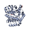
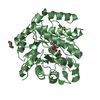
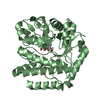
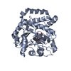
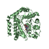
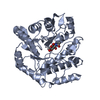
| Title | GlcNAc-inspired cyclophellitol bound to NagZ | ||||||
 Components Components | Beta-hexosaminidase Hexosaminidase Hexosaminidase | ||||||
 Keywords Keywords | HYDROLASE / Antibiotic potentiator / Antibiotic adjuvant / NagZ / glycosides / epoxide / GlcNAc / Antibiotic resistance / AmpC | ||||||
| Function / homology |  Function and homology informationbeta-N-acetylhexosaminidase activity / beta-N-acetylhexosaminidase / peptidoglycan turnover / N-acetyl-beta-D-galactosaminidase activity / peptidoglycan biosynthetic process / cell wall organization / regulation of cell shape / carbohydrate metabolic process / cell cycle / cell division / cytoplasm Function and homology informationbeta-N-acetylhexosaminidase activity / beta-N-acetylhexosaminidase / peptidoglycan turnover / N-acetyl-beta-D-galactosaminidase activity / peptidoglycan biosynthetic process / cell wall organization / regulation of cell shape / carbohydrate metabolic process / cell cycle / cell division / cytoplasmSimilarity search - Function | ||||||
| Biological species |  Burkholderia cenocepacia (bacteria) Burkholderia cenocepacia (bacteria) | ||||||
| Method | X-RAY DIFFRACTION / MOLECULAR REPLACEMENT / Resolution: 1.929 Å | ||||||
 Authors Authors | Mark, B.L. / Winogrodzki, J.L. | ||||||
| Funding support |  Canada, 1items Canada, 1items
| ||||||
 Citation Citation | Journal: Chem. Commun. (Camb.) / Year: 2018 Title: A mechanism-based GlcNAc-inspired cyclophellitol inactivator of the peptidoglycan recycling enzyme NagZ reverses resistance to beta-lactams in Pseudomonas aeruginosa. Authors: Ho, L.A. / Winogrodzki, J.L. / Debowski, A.W. / Madden, Z. / Vocadlo, D.J. / Mark, B.L. / Stubbs, K.A. | ||||||
| History |
|
- Structure visualization
| Structure viewer | Molecule: MolmilJmol/JSmol |
|---|
- Downloads & links
-Download
| PDBx/mmCIF format | 6dte.cif.gz | 270.2 KB | Display | PDBx/mmCIF format |
|---|---|---|---|---|
| PDB format | pdb6dte.ent.gz | 217.6 KB | Display | PDB format |
| PDBx/mmJSON format | 6dte.json.gz | Tree view | PDBx/mmJSON format | |
| Others |  Other downloads Other downloads |
-Validation report
| Arichive directory | https://data.pdbj.org/pub/pdb/validation_reports/dt/6dteftp://data.pdbj.org/pub/pdb/validation_reports/dt/6dte | HTTPS FTP |
|---|
-Related structure data
| Related structure data | 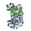 4g6cS S: Starting model for refinement |
|---|---|
| Similar structure data |


-Links
 PDBj
PDBj- Assembly
| Deposited unit | 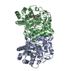
| ||||||||
|---|---|---|---|---|---|---|---|---|---|
| 1 | 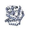
| ||||||||
| 2 | 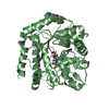
| ||||||||
| Unit cell |
|
-Components
| #1: Protein | Hexosaminidase / Beta-N-acetylhexosaminidase / N-acetyl-beta-glucosaminidase Mass: 38116.766 Da / Num. of mol.: 2 Source method: isolated from a genetically manipulated source Source: (gene. exp.) Burkholderia cenocepacia (bacteria)Gene: nagZ, A8E72_12990, A8F51_10310, A8F55_30255, BCN122_I0864, BCN122_I2678, BCN122_II1911, UE95_02480 Production host: Escherichia coli BL21(DE3) (bacteria) / Strain (production host): BL21(DE3)References: UniProt: A0A125HFC0, beta-N-acetylhexosaminidase#2: Chemical |   Mass: 289.206 Da / Num. of mol.: 2 / Source method: isolated from a natural source / Formula: C9H14F3NO6 Mass: 289.206 Da / Num. of mol.: 2 / Source method: isolated from a natural source / Formula: C9H14F3NO6#3: Water | ChemComp-HOH / | Water Mass: 18.015 Da / Num. of mol.: 843 / Source method: isolated from a natural source / Formula: H2O Mass: 18.015 Da / Num. of mol.: 843 / Source method: isolated from a natural source / Formula: H2O |
|---|
-Experimental details
-Experiment
| Experiment | Method: X-RAY DIFFRACTION / Number of used crystals: 1 |
|---|
- Sample preparation
| Crystal | Density Matthews: 1.93 Å3/Da / Density % sol: 36.43 % |
|---|---|
| Crystal grow | Temperature: 298 K / Method: vapor diffusion, hanging drop / Details: 30-32% PEG8000, 0.1M MES pH 6.6-6.8 / PH range: 6.6-6.8 |
-Data collection
| Diffraction | Mean temperature: 100 K |
|---|---|
| Diffraction source | Source: ROTATING ANODE / Type: RIGAKU MICROMAX-007 HF / Wavelength: 1.54 Å |
| Detector | Type: RIGAKU RAXIS IV++ / Detector: IMAGE PLATE / Date: Dec 4, 2017 |
| Radiation | Protocol: SINGLE WAVELENGTH / Monochromatic (M) / Laue (L): M / Scattering type: x-ray |
| Radiation wavelength | Wavelength: 1.54 Å / Relative weight: 1 |
| Reflection | Resolution: 1.929→67.27 Å / Num. obs: 42981 / % possible obs: 99.9 % / Redundancy: 3.6 % / Biso Wilson estimate: 14.87 Å2 / CC1/2: 0.993 / Rmerge(I) obs: 0.082 / Net I/σ(I): 9.3 |
| Reflection shell | Resolution: 1.929→1.97 Å / Redundancy: 3.3 % / Rmerge(I) obs: 0.309 / Mean I/σ(I) obs: 3.3 / Num. unique obs: 2882 / CC1/2: 0.833 / % possible all: 98.9 |
- Processing
| Software |
| |||||||||||||||||||||||||||||||||||||||||||||||||||||||||||||||||||||||||||||||||||||||||||||||||||||||||
|---|---|---|---|---|---|---|---|---|---|---|---|---|---|---|---|---|---|---|---|---|---|---|---|---|---|---|---|---|---|---|---|---|---|---|---|---|---|---|---|---|---|---|---|---|---|---|---|---|---|---|---|---|---|---|---|---|---|---|---|---|---|---|---|---|---|---|---|---|---|---|---|---|---|---|---|---|---|---|---|---|---|---|---|---|---|---|---|---|---|---|---|---|---|---|---|---|---|---|---|---|---|---|---|---|---|---|
| Refinement | Method to determine structure: MOLECULAR REPLACEMENT Starting model: 4G6C Resolution: 1.929→40.226 Å / SU ML: 0.2 / Cross valid method: FREE R-VALUE / σ(F): 1.34 / Phase error: 19.16 / Stereochemistry target values: ML
| |||||||||||||||||||||||||||||||||||||||||||||||||||||||||||||||||||||||||||||||||||||||||||||||||||||||||
| Solvent computation | Shrinkage radii: 0.9 Å / VDW probe radii: 1.11 Å / Solvent model: FLAT BULK SOLVENT MODEL | |||||||||||||||||||||||||||||||||||||||||||||||||||||||||||||||||||||||||||||||||||||||||||||||||||||||||
| Refinement step | Cycle: LAST / Resolution: 1.929→40.226 Å
| |||||||||||||||||||||||||||||||||||||||||||||||||||||||||||||||||||||||||||||||||||||||||||||||||||||||||
| Refine LS restraints |
| |||||||||||||||||||||||||||||||||||||||||||||||||||||||||||||||||||||||||||||||||||||||||||||||||||||||||
| LS refinement shell |
|
