 Movie
Movie Controller


+ Open data

- Basic information
| Entry | Database: PDB / ID: 6dk3 | |||||||||
|---|---|---|---|---|---|---|---|---|---|---|
| Title | HUMAN MITOCHONDRIAL SERINE HYDROXYMETHYLTRANSFERASe 2 | |||||||||
 Components Components | Serine hydroxymethyltransferase, mitochondrial | |||||||||
 Keywords Keywords | TRANSFERASE / SHMT2 / Structural Genomics / Structural Genomics Consortium / SGC | |||||||||
| Function / homology |  Function and homology information Function and homology informationBRISC complex / L-allo-threonine aldolase activity / regulation of mitochondrial translation / purine nucleobase biosynthetic process / L-serine metabolic process / glycine metabolic process / L-serine biosynthetic process / serine binding / glycine hydroxymethyltransferase / glycine hydroxymethyltransferase activity ...BRISC complex / L-allo-threonine aldolase activity / regulation of mitochondrial translation / purine nucleobase biosynthetic process / L-serine metabolic process / glycine metabolic process / L-serine biosynthetic process / serine binding / glycine hydroxymethyltransferase / glycine hydroxymethyltransferase activity / glycine biosynthetic process from serine / L-serine catabolic process / Metabolism of folate and pterines / regulation of oxidative phosphorylation / tetrahydrofolate metabolic process / response to type I interferon / protein K63-linked deubiquitination / tetrahydrofolate interconversion / regulation of aerobic respiration / mitochondrial nucleoid / folic acid metabolic process / RHOG GTPase cycle / mRNA regulatory element binding translation repressor activity / protein tetramerization / mRNA 5'-UTR binding / microtubule cytoskeleton / pyridoxal phosphate binding / one-carbon metabolic process / protein homotetramerization / mitochondrial inner membrane / mitochondrial matrix / chromatin binding / positive regulation of cell population proliferation / protein homodimerization activity / mitochondrion / extracellular exosome / zinc ion binding / nucleus / cytosol / cytoplasmSimilarity search - Function | |||||||||
| Biological species |  Homo sapiens (human) Homo sapiens (human) | |||||||||
| Method | X-RAY DIFFRACTION / SYNCHROTRON / MOLECULAR REPLACEMENT / molecular replacement / Resolution: 2.04 Å | |||||||||
 Authors Authors | Dong, A. / Wu, H. / Zeng, H. / Loppnau, P. / Sundstrom, M. / Arrowsmith, C.H. / Edwards, A.M. / Bochkarev, A. / Plotnikov, A.N. / Structural Genomics Consortium (SGC) | |||||||||
 Citation Citation | Journal: to be published Title: HUMAN MITOCHONDRIAL SERINE HYDROXYMETHYLTRANSFERASe 2 Authors: Wu, H. / Dong, A. / Zeng, H. / Loppnau, P. / Sundstrom, M. / Arrowsmith, C.H. / Edwards, A.M. / Bochkarev, A. / Plotnikov, A.N. / Structural Genomics Consortium (SGC) | |||||||||
| History |
|
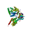
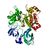
- Structure visualization
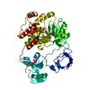
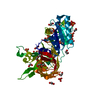
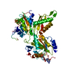
| Structure viewer | Molecule: MolmilJmol/JSmol |
|---|
- Downloads & links
-Download
| PDBx/mmCIF format | 6dk3.cif.gz | 101.3 KB | Display | PDBx/mmCIF format |
|---|---|---|---|---|
| PDB format | pdb6dk3.ent.gz | 73.4 KB | Display | PDB format |
| PDBx/mmJSON format | 6dk3.json.gz | Tree view | PDBx/mmJSON format | |
| Others |  Other downloads Other downloads |
-Validation report
| Arichive directory | https://data.pdbj.org/pub/pdb/validation_reports/dk/6dk3ftp://data.pdbj.org/pub/pdb/validation_reports/dk/6dk3 | HTTPS FTP |
|---|
-Related structure data
| Related structure data |  1ls3S S: Starting model for refinement |
|---|---|
| Similar structure data |


-Links
 PDBj
PDBj

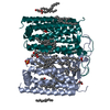
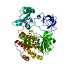
- Assembly
| Deposited unit | 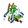
| ||||||||
|---|---|---|---|---|---|---|---|---|---|
| 1 |
| ||||||||
| 2 | 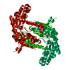
| ||||||||
| Unit cell |
| ||||||||
| Components on special symmetry positions |
|
-Components
| #1: Protein | / SHMT / Glycine hydroxymethyltransferase / Serine methylase Mass: 54111.504 Da / Num. of mol.: 1 Source method: isolated from a genetically manipulated source Source: (gene. exp.) Homo sapiens (human) / Gene: SHMT2 / Plasmid: PET28 / Production host:  Escherichia coli (E. coli) / Strain (production host): BL21(DE3) Escherichia coli (E. coli) / Strain (production host): BL21(DE3)References: UniProt: P34897, glycine hydroxymethyltransferase | ||
|---|---|---|---|
| #2: Chemical | ChemComp-UNX /   Num. of mol.: 6 / Source method: obtained synthetically Num. of mol.: 6 / Source method: obtained synthetically#3: Water | ChemComp-HOH / | Water Mass: 18.015 Da / Num. of mol.: 167 / Source method: isolated from a natural source / Formula: H2O Mass: 18.015 Da / Num. of mol.: 167 / Source method: isolated from a natural source / Formula: H2O |
-Experimental details
-Experiment
| Experiment | Method: X-RAY DIFFRACTION / Number of used crystals: 1 |
|---|
- Sample preparation
| Crystal | Density Matthews: 2.35 Å3/Da / Density % sol: 47.59 % |
|---|---|
| Crystal grow | Temperature: 297 K / Method: vapor diffusion, hanging drop / pH: 8 / Details: 5% PEG 8000, 0.1 M Tris-HCL pH8.0 |
-Data collection
| Diffraction | Mean temperature: 100 K | |||||||||||||||||||||||||||||||||||||||||||||||||||||||||||||||||||||||||||||||||||||||||||||||||||||||||||||||||||||||||||||||||||||||||||||||||||
|---|---|---|---|---|---|---|---|---|---|---|---|---|---|---|---|---|---|---|---|---|---|---|---|---|---|---|---|---|---|---|---|---|---|---|---|---|---|---|---|---|---|---|---|---|---|---|---|---|---|---|---|---|---|---|---|---|---|---|---|---|---|---|---|---|---|---|---|---|---|---|---|---|---|---|---|---|---|---|---|---|---|---|---|---|---|---|---|---|---|---|---|---|---|---|---|---|---|---|---|---|---|---|---|---|---|---|---|---|---|---|---|---|---|---|---|---|---|---|---|---|---|---|---|---|---|---|---|---|---|---|---|---|---|---|---|---|---|---|---|---|---|---|---|---|---|---|---|---|
| Diffraction source | Source: SYNCHROTRON / Site: CHESS  / Beamline: F1 / Wavelength: 0.912386 Å / Beamline: F1 / Wavelength: 0.912386 Å | |||||||||||||||||||||||||||||||||||||||||||||||||||||||||||||||||||||||||||||||||||||||||||||||||||||||||||||||||||||||||||||||||||||||||||||||||||
| Detector | Type: ADSC QUANTUM 4 / Detector: CCD / Date: May 8, 2005 | |||||||||||||||||||||||||||||||||||||||||||||||||||||||||||||||||||||||||||||||||||||||||||||||||||||||||||||||||||||||||||||||||||||||||||||||||||
| Radiation | Protocol: SINGLE WAVELENGTH / Monochromatic (M) / Laue (L): M / Scattering type: x-ray | |||||||||||||||||||||||||||||||||||||||||||||||||||||||||||||||||||||||||||||||||||||||||||||||||||||||||||||||||||||||||||||||||||||||||||||||||||
| Radiation wavelength | Wavelength: 0.912386 Å / Relative weight: 1 | |||||||||||||||||||||||||||||||||||||||||||||||||||||||||||||||||||||||||||||||||||||||||||||||||||||||||||||||||||||||||||||||||||||||||||||||||||
| Reflection | Resolution: 2.04→50 Å / Num. obs: 31273 / % possible obs: 98.6 % / Redundancy: 3.5 % / Rmerge(I) obs: 0.06 / Χ2: 1.354 / Net I/σ(I): 14.3 / Num. measured all: 109928 | |||||||||||||||||||||||||||||||||||||||||||||||||||||||||||||||||||||||||||||||||||||||||||||||||||||||||||||||||||||||||||||||||||||||||||||||||||
| Reflection shell |
|
-Phasing
| Phasing | Method: molecular replacement |
|---|
- Processing
| Software |
| ||||||||||||||||||||||||||||||||||||||||||||||||||||||||||||
|---|---|---|---|---|---|---|---|---|---|---|---|---|---|---|---|---|---|---|---|---|---|---|---|---|---|---|---|---|---|---|---|---|---|---|---|---|---|---|---|---|---|---|---|---|---|---|---|---|---|---|---|---|---|---|---|---|---|---|---|---|---|
| Refinement | Method to determine structure: MOLECULAR REPLACEMENT Starting model: 1LS3 Resolution: 2.04→33.56 Å / Cor.coef. Fo:Fc: 0.957 / Cor.coef. Fo:Fc free: 0.933 / SU B: 5.363 / SU ML: 0.143 / Cross valid method: THROUGHOUT / σ(F): 0 / ESU R: 0.181 / ESU R Free: 0.177 / Stereochemistry target values: MAXIMUM LIKELIHOOD Details: HYDROGENS HAVE BEEN ADDED IN THE RIDING POSITIONS U VALUES : REFINED INDIVIDUALLY
| ||||||||||||||||||||||||||||||||||||||||||||||||||||||||||||
| Solvent computation | Ion probe radii: 0.8 Å / Shrinkage radii: 0.8 Å / VDW probe radii: 1.2 Å / Solvent model: MASK | ||||||||||||||||||||||||||||||||||||||||||||||||||||||||||||
| Displacement parameters | Biso max: 96.04 Å2 / Biso mean: 45.721 Å2 / Biso min: 28.21 Å2
| ||||||||||||||||||||||||||||||||||||||||||||||||||||||||||||
| Refinement step | Cycle: final / Resolution: 2.04→33.56 Å
| ||||||||||||||||||||||||||||||||||||||||||||||||||||||||||||
| Refine LS restraints |
| ||||||||||||||||||||||||||||||||||||||||||||||||||||||||||||
| LS refinement shell | Resolution: 2.043→2.096 Å / Rfactor Rfree error: 0 / Total num. of bins used: 20
|
