 Movie
Movie Controller


+ Open data

- Basic information
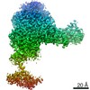
| Entry | Database: PDB / ID: 6dk0 | |||||||||
|---|---|---|---|---|---|---|---|---|---|---|
| Title | Human sigma-1 receptor bound to NE-100 | |||||||||
 Components Components | Sigma non-opioid intracellular receptor 1 | |||||||||
 Keywords Keywords |  MEMBRANE PROTEIN / Antagonist bound / Receptor / sigma-1 receptor MEMBRANE PROTEIN / Antagonist bound / Receptor / sigma-1 receptor | |||||||||
| Function / homology |  Function and homology information Function and homology informationG protein-coupled opioid receptor activity / nuclear outer membrane / anchoring junction / lipid transport / nuclear inner membrane / protein homotrimerization / regulation of neuron apoptotic process / lipid droplet / postsynaptic density membrane / nuclear envelope ...G protein-coupled opioid receptor activity / nuclear outer membrane / anchoring junction / lipid transport / nuclear inner membrane / protein homotrimerization / regulation of neuron apoptotic process / lipid droplet / postsynaptic density membrane / nuclear envelope / nervous system development / growth cone / cytoplasmic vesicle / Potential therapeutics for SARS / postsynaptic density / endoplasmic reticulum membrane / endoplasmic reticulum / membrane / identical protein binding / cytosolSimilarity search - Function | |||||||||
| Biological species |  Homo sapiens (human) Homo sapiens (human) | |||||||||
| Method | X-RAY DIFFRACTION / SYNCHROTRON / MOLECULAR REPLACEMENT / Resolution: 2.9 Å | |||||||||
 Authors Authors | Schmidt, H.R. / Kruse, A.C. | |||||||||
| Funding support |  United States, 2items United States, 2items
| |||||||||
 Citation Citation | Journal: Nat. Struct. Mol. Biol. / Year: 2018 Title: Structural basis for sigma1receptor ligand recognition. Authors: Schmidt, H.R. / Betz, R.M. / Dror, R.O. / Kruse, A.C. | |||||||||
| History |
|
- Structure visualization
| Structure viewer | Molecule: MolmilJmol/JSmol |
|---|
- Downloads & links
-Download
| PDBx/mmCIF format | 6dk0.cif.gz | 151.2 KB | Display | PDBx/mmCIF format |
|---|---|---|---|---|
| PDB format | pdb6dk0.ent.gz | 116.4 KB | Display | PDB format |
| PDBx/mmJSON format | 6dk0.json.gz | Tree view | PDBx/mmJSON format | |
| Others |  Other downloads Other downloads |
-Validation report
| Arichive directory | https://data.pdbj.org/pub/pdb/validation_reports/dk/6dk0ftp://data.pdbj.org/pub/pdb/validation_reports/dk/6dk0 | HTTPS FTP |
|---|
-Related structure data
| Related structure data |  6djzC  6dk1C  5hk1S S: Starting model for refinement C: citing same article ( |
|---|---|
| Similar structure data |


-Links
 PDBj
PDBj


- Assembly
| Deposited unit | 
| ||||||||
|---|---|---|---|---|---|---|---|---|---|
| 1 |
| ||||||||
| Unit cell |
| ||||||||
| Components on special symmetry positions |
|
-Components
-Protein , 1 types, 3 molecules ABC
| #1: Protein | Mass: 25447.879 Da / Num. of mol.: 3 Source method: isolated from a genetically manipulated source Source: (gene. exp.) Homo sapiens (human) / Gene: SIGMAR1, OPRS1, SRBP, AAG8 / Production host:   Spodoptera frugiperda (fall armyworm) / References: UniProt: Q99720 Spodoptera frugiperda (fall armyworm) / References: UniProt: Q99720 |
|---|
-Non-polymers , 5 types, 124 molecules 


| #2: Chemical | NE-100 Mass: 355.514 Da / Num. of mol.: 3 / Source method: obtained synthetically / Formula: C23H33NO2 / Comment: antagonist*YM Mass: 355.514 Da / Num. of mol.: 3 / Source method: obtained synthetically / Formula: C23H33NO2 / Comment: antagonist*YM#3: Chemical | ChemComp-SO4 / Sulfate Mass: 96.063 Da / Num. of mol.: 14 / Source method: obtained synthetically / Formula: SO4 Mass: 96.063 Da / Num. of mol.: 14 / Source method: obtained synthetically / Formula: SO4#4: Chemical | Glycerol Mass: 92.094 Da / Num. of mol.: 3 / Source method: obtained synthetically / Formula: C3H8O3 Mass: 92.094 Da / Num. of mol.: 3 / Source method: obtained synthetically / Formula: C3H8O3#5: Chemical | ChemComp-OLC / (  Mass: 356.540 Da / Num. of mol.: 8 / Source method: obtained synthetically / Formula: C21H40O4 Mass: 356.540 Da / Num. of mol.: 8 / Source method: obtained synthetically / Formula: C21H40O4#6: Water | ChemComp-HOH / | WaterMass: 18.015 Da / Num. of mol.: 96 / Source method: isolated from a natural source / Formula: H2O |
|---|
-Experimental details
-Experiment
| Experiment | Method: X-RAY DIFFRACTION / Number of used crystals: 1 |
|---|
- Sample preparation
| Crystal | Density Matthews: 3.87 Å3/Da / Density % sol: 68.25 % |
|---|---|
| Crystal grow | Temperature: 293 K / Method: lipidic cubic phase Details: Protein was reconstituted in cubic phase by mixing with a 1:1.5 (w:w) protein:lipid ratio. Lipid used was a 10:1 (w:w) mix of monoolein with cholesterol. Crystals were grown in 400-500 mM ...Details: Protein was reconstituted in cubic phase by mixing with a 1:1.5 (w:w) protein:lipid ratio. Lipid used was a 10:1 (w:w) mix of monoolein with cholesterol. Crystals were grown in 400-500 mM lithium sulfate, 30-40% PEG 300, 1% hexanediol, 0.1 M MES pH 5.8-6.0 PH range: 5.8-6.0 |
-Data collection
| Diffraction | Mean temperature: 80 K |
|---|---|
| Diffraction source | Source: SYNCHROTRON / Site: APS / Beamline: 23-ID-B / Wavelength: 1.033 Å |
| Detector | Type: DECTRIS EIGER X 16M / Detector: PIXEL / Date: Jun 8, 2017 |
| Radiation | Monochromator: Si(111) / Protocol: SINGLE WAVELENGTH / Monochromatic (M) / Laue (L): M / Scattering type: x-ray |
| Radiation wavelength | Wavelength: 1.033 Å / Relative weight: 1 |
| Reflection | Resolution: 2.9→50 Å / Num. obs: 25810 / % possible obs: 95.3 % / Redundancy: 7.2 % / CC1/2: 0.989 / Net I/σ(I): 5.6 |
| Reflection shell | Resolution: 2.9→3 Å / Mean I/σ(I) obs: 0.5 / CC1/2: 0.24 / % possible all: 96.8 |
- Processing
| Software |
| ||||||||||||||||||||||||||||||||||||||||||||||||||||||||||||||||||||||||||||||||||||||||||||||||||||||||||||||||
|---|---|---|---|---|---|---|---|---|---|---|---|---|---|---|---|---|---|---|---|---|---|---|---|---|---|---|---|---|---|---|---|---|---|---|---|---|---|---|---|---|---|---|---|---|---|---|---|---|---|---|---|---|---|---|---|---|---|---|---|---|---|---|---|---|---|---|---|---|---|---|---|---|---|---|---|---|---|---|---|---|---|---|---|---|---|---|---|---|---|---|---|---|---|---|---|---|---|---|---|---|---|---|---|---|---|---|---|---|---|---|---|---|---|
| Refinement | Method to determine structure: MOLECULAR REPLACEMENT Starting model: 5HK1 Resolution: 2.9→46.176 Å / SU ML: 0.5 / Cross valid method: FREE R-VALUE / σ(F): 1.33 / Phase error: 31.85
| ||||||||||||||||||||||||||||||||||||||||||||||||||||||||||||||||||||||||||||||||||||||||||||||||||||||||||||||||
| Solvent computation | Shrinkage radii: 0.9 Å / VDW probe radii: 1.11 Å | ||||||||||||||||||||||||||||||||||||||||||||||||||||||||||||||||||||||||||||||||||||||||||||||||||||||||||||||||
| Refinement step | Cycle: LAST / Resolution: 2.9→46.176 Å
| ||||||||||||||||||||||||||||||||||||||||||||||||||||||||||||||||||||||||||||||||||||||||||||||||||||||||||||||||
| Refine LS restraints |
| ||||||||||||||||||||||||||||||||||||||||||||||||||||||||||||||||||||||||||||||||||||||||||||||||||||||||||||||||
| LS refinement shell |
|
