[English] 日本語
Yorodumi
- PDB-6d6y: AprA Methyltransferase 2 - GNAT didomain in complex with SAH -

+
Open data


ID or keywords:

Loading...

-
Basic information

Entry
Database: PDB / ID: 6d6y
TitleAprA Methyltransferase 2 - GNAT didomain in complex with SAH
ComponentsAprA Methyltransferase 2
KeywordsTRANSFERASE / methyltransferase / decarboxylase / apratoxin / GCN5 related N-acetyltransferase
Function / homology
Function and homology information


phosphopantetheine binding / transferase activity
Similarity search - Function
Methyltransferase type 12 / Methyltransferase domain / Polyketide synthase, phosphopantetheine-binding domain / Phosphopantetheine attachment site / Phosphopantetheine attachment site / ACP-like superfamily / Carrier protein (CP) domain profile. / Phosphopantetheine binding ACP domain / Prokaryotic membrane lipoprotein lipid attachment site profile. / Winged helix-like DNA-binding domain superfamily / S-adenosyl-L-methionine-dependent methyltransferase superfamily
Similarity search - Domain/homology
S-ADENOSYL-L-HOMOCYSTEINE / trimethylamine oxide / Carrier domain-containing protein
Similarity search - Component
Biological speciesMoorea bouillonii (bacteria)
MethodX-RAY DIFFRACTION / SYNCHROTRON / MOLECULAR REPLACEMENT / Resolution: 2.246 Å
AuthorsSikkema, A.P. / Smith, J.L.
Funding support United States, 3items
OrganizationGrant numberCountry
National Institutes of Health/National Institute of Diabetes and Digestive and Kidney Disease (NIH/NIDDK)DK042303 United States
National Institutes of Health/National Cancer Institute (NIH/NCI)CA108874 United States
National Institutes of Health/National Institute of General Medical Sciences (NIH/NIGMS)GM008270 United States
Citation
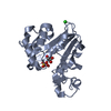
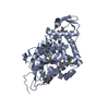
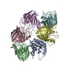
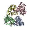
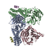
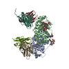
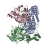
Journal: ACS Chem Biol / Year: 2018
Title: Biosynthesis of t-Butyl in Apratoxin A: Functional Analysis and Architecture of a PKS Loading Module.
Authors: Meredith A Skiba / Andrew P Sikkema / Nathan A Moss / Andrew N Lowell / Min Su / Rebecca M Sturgis / Lena Gerwick / William H Gerwick / David H Sherman / Janet L Smith /
Abstract: The unusual feature of a t-butyl group is found in several marine-derived natural products including apratoxin A, a Sec61 inhibitor produced by the cyanobacterium Moorea bouillonii PNG 5-198. Here, ...The unusual feature of a t-butyl group is found in several marine-derived natural products including apratoxin A, a Sec61 inhibitor produced by the cyanobacterium Moorea bouillonii PNG 5-198. Here, we determine that the apratoxin A t-butyl group is formed as a pivaloyl acyl carrier protein (ACP) by AprA, the polyketide synthase (PKS) loading module of the apratoxin A biosynthetic pathway. AprA contains an inactive "pseudo" GCN5-related N-acetyltransferase domain (ΨGNAT) flanked by two methyltransferase domains (MT1 and MT2) that differ distinctly in sequence. Structural, biochemical, and precursor incorporation studies reveal that MT2 catalyzes unusually coupled decarboxylation and methylation reactions to transform dimethylmalonyl-ACP, the product of MT1, to pivaloyl-ACP. Further, pivaloyl-ACP synthesis is primed by the fatty acid synthase malonyl acyltransferase (FabD), which compensates for the ΨGNAT and provides the initial acyl-transfer step to form AprA malonyl-ACP. Additionally, images of AprA from negative stain electron microscopy reveal multiple conformations that may facilitate the individual catalytic steps of the multienzyme module.
History
DepositionApr 23, 2018Deposition site: RCSB / Processing site: RCSB
Revision 1.0May 9, 2018Provider: repository / Type: Initial release
Revision 1.1Jun 27, 2018Group: Data collection / Database references / Category: citation
Item: _citation.journal_volume / _citation.page_first / _citation.page_last
Revision 1.2Dec 4, 2019Group: Author supporting evidence / Category: pdbx_audit_support / Item: _pdbx_audit_support.funding_organization
Revision 1.3Oct 4, 2023Group: Data collection / Database references / Refinement description
Category: chem_comp_atom / chem_comp_bond ...chem_comp_atom / chem_comp_bond / database_2 / pdbx_initial_refinement_model
Item: _database_2.pdbx_DOI / _database_2.pdbx_database_accession

-
Structure visualization

Structure viewerMolecule:
MolmilJmol/JSmol

Downloads & links

-
Assembly

Deposited unit
A: AprA Methyltransferase 2
hetero molecules


Theoretical massNumber of molelcules
Total (without water)60,5903
Polymers60,1311
Non-polymers4602
Water2,252125
1
A: AprA Methyltransferase 2
hetero molecules

A: AprA Methyltransferase 2
hetero molecules


Theoretical massNumber of molelcules
Total (without water)121,1816
Polymers120,2622
Non-polymers9194
Water362
TypeNameSymmetry operationNumber
identity operation1_555x,y,z1
crystal symmetry operation2_656-x+1,y,-z+11
2


  • Idetical with deposited unit
  • defined by software
TypeNameSymmetry operationNumber
identity operation1_555x,y,z1
Buried area0 Å2
ΔGint0 kcal/mol
Surface area22980 Å2
MethodPISA
Unit cell
Length a, b, c (Å)151.395, 54.206, 109.300
Angle α, β, γ (deg.)90.000, 131.810, 90.000
Int Tables number5
Space group name H-MC121

-
Components

#1: Protein AprA Methyltransferase 2


Mass: 60130.934 Da / Num. of mol.: 1 / Fragment: GNAT didomain (UNP residues 503-1022)
Source method: isolated from a genetically manipulated source
Source: (gene. exp.) Moorea bouillonii (bacteria) / Gene: BJP37_16135 / Plasmid: pMCSG7 / Production host: Escherichia coli BL21(DE3) (bacteria)
References: UniProt: A0A1U7N2Z8, Transferases; Transferring one-carbon groups; Methyltransferases
#2: Chemical ChemComp-TMO / trimethylamine oxide / Trimethylamine N-oxide


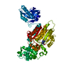
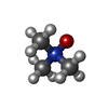
Mass: 75.110 Da / Num. of mol.: 1 / Source method: obtained synthetically / Formula: C3H9NO
#3: Chemical ChemComp-SAH / S-ADENOSYL-L-HOMOCYSTEINE / S-Adenosyl-L-homocysteine


Type: L-peptide linking / Mass: 384.411 Da / Num. of mol.: 1 / Source method: obtained synthetically / Formula: C14H20N6O5S / Feature type: SUBJECT OF INVESTIGATION
#4: Water ChemComp-HOH / water / Water


Mass: 18.015 Da / Num. of mol.: 125 / Source method: isolated from a natural source / Formula: H2O

-
Experimental details

-
Experiment

ExperimentMethod: X-RAY DIFFRACTION / Number of used crystals: 1

-
Sample preparation

CrystalDensity Matthews: 2.78 Å3/Da / Density % sol: 55.75 %
Crystal growTemperature: 293 K / Method: vapor diffusion, sitting drop / pH: 7.5
Details: 0.01-0.05 M trimethylamine N-oxide, 12-17% PEG8000, 0.12 M Tris, pH 7.5

-
Data collection

DiffractionMean temperature: 100 K
Diffraction sourceSource: SYNCHROTRON / Site: APS / Beamline: 23-ID-B / Wavelength: 1.0332 Å
DetectorType: DECTRIS EIGER X 16M / Detector: PIXEL / Date: Nov 9, 2016
RadiationMonochromator: Double crystal cryo-cooled Si(111) / Protocol: SINGLE WAVELENGTH / Monochromatic (M) / Laue (L): M / Scattering type: x-ray
Radiation wavelengthWavelength: 1.0332 Å / Relative weight: 1
ReflectionResolution: 2.246→48.861 Å / Num. obs: 60876 / % possible obs: 98.4 % / Redundancy: 6.067 % / Biso Wilson estimate: 52.96 Å2 / CC1/2: 0.998 / Rmerge(I) obs: 0.09 / Rrim(I) all: 0.099 / Χ2: 1.039 / Net I/σ(I): 12.09
Reflection shell

Diffraction-ID: 1

Resolution (Å)Redundancy (%)Rmerge(I) obsMean I/σ(I) obsNum. unique obsCC1/2Rrim(I) all% possible all
2.246-2.386.3560.9981.650170.721.08697.8
2.38-2.556.0980.7122.4547530.7930.77998.8
2.55-2.755.8340.4214.1243900.9230.46297.7
2.75-3.016.1760.2397.4941050.9740.26199
3.01-3.366.1110.13713.137300.9910.1599.1
3.36-3.885.7860.08920.7432670.9950.09897.8
3.88-4.756.2350.05829.7728010.9980.06399.2
4.75-6.685.9860.05330.6122030.9980.05898.8
6.68-48.8615.6270.03936.2212460.9990.04396

-
Processing

Software
NameVersionClassification
XDSdata reduction
XSCALEdata scaling
PHENIX1.10.1refinement
PDB_EXTRACT3.24data extraction
PHENIX1.10.1phasing
RefinementMethod to determine structure: MOLECULAR REPLACEMENT
Starting model: PDB entry 5THY & 2REE
Resolution: 2.246→48.861 Å / SU ML: 0.34 / Cross valid method: THROUGHOUT / σ(F): 1.36 / Phase error: 25.51
RfactorNum. reflection% reflection
Rfree0.2305 3063 5.03 %
Rwork0.1742 --
obs0.177 60876 98.49 %
Solvent computationShrinkage radii: 0.9 Å / VDW probe radii: 1.11 Å
Displacement parametersBiso max: 179.05 Å2 / Biso mean: 68.1807 Å2 / Biso min: 29.4 Å2
Refinement stepCycle: final / Resolution: 2.246→48.861 Å
ProteinNucleic acidLigandSolventTotal
Num. atoms4147 0 31 125 4303
Biso mean--96.21 62.18 -
Num. residues----511
Refine LS restraints
Refine-IDTypeDev idealNumber
X-RAY DIFFRACTIONf_bond_d0.0084290
X-RAY DIFFRACTIONf_angle_d0.9835817
X-RAY DIFFRACTIONf_chiral_restr0.055651
X-RAY DIFFRACTIONf_plane_restr0.006733
X-RAY DIFFRACTIONf_dihedral_angle_d19.9731574
LS refinement shell

Refine-ID: X-RAY DIFFRACTION / Rfactor Rfree error: 0 / Total num. of bins used: 22

Resolution (Å)Rfactor RfreeNum. reflection RfreeRfactor RworkNum. reflection RworkNum. reflection all% reflection obs (%)
2.246-2.28110.32071280.28772562269096
2.2811-2.31850.36751510.294726722823100
2.3185-2.35850.33991400.29762627276797
2.3585-2.40130.32061400.291225972737100
2.4013-2.44750.35751230.26842683280699
2.4475-2.49750.29461450.25462560270598
2.4975-2.55180.31791470.257226412788100
2.5518-2.61110.30311460.25812608275497
2.6111-2.67640.28081320.25642601273396
2.6764-2.74880.35761250.24542606273199
2.7488-2.82970.34461630.238626152778100
2.8297-2.9210.26991220.22232684280699
2.921-3.02540.24831610.2192605276699
3.0254-3.14650.27641290.201627042833100
3.1465-3.28970.23121510.18262620277199
3.2897-3.46310.20811390.1692641278099
3.4631-3.680.23541250.16262684280999
3.68-3.9640.21491320.14732530266296
3.964-4.36270.17671390.131726542793100
4.3627-4.99340.18561470.125826782825100
4.9934-6.28910.22631380.148426392777100
6.2891-48.87210.16991400.1332602274297

+
About Yorodumi

-
News

-
Feb 9, 2022. New format data for meta-information of EMDB entries

New format data for meta-information of EMDB entries

  • Version 3 of the EMDB header file is now the official format.
  • The previous official version 1.9 will be removed from the archive.

Related info.:EMDB header

External links:wwPDB to switch to version 3 of the EMDB data model

-
Aug 12, 2020. Covid-19 info

Covid-19 info

URL: https://pdbj.org/emnavi/covid19.php

New page: Covid-19 featured information page in EM Navigator.

Related info.:Covid-19 info / Mar 5, 2020. Novel coronavirus structure data

+
Mar 5, 2020. Novel coronavirus structure data

Novel coronavirus structure data

Related info.:Yorodumi Speices / Aug 12, 2020. Covid-19 info

External links:COVID-19 featured content - PDBj / Molecule of the Month (242):Coronavirus Proteases

+
Jan 31, 2019. EMDB accession codes are about to change! (news from PDBe EMDB page)

EMDB accession codes are about to change! (news from PDBe EMDB page)

  • The allocation of 4 digits for EMDB accession codes will soon come to an end. Whilst these codes will remain in use, new EMDB accession codes will include an additional digit and will expand incrementally as the available range of codes is exhausted. The current 4-digit format prefixed with “EMD-” (i.e. EMD-XXXX) will advance to a 5-digit format (i.e. EMD-XXXXX), and so on. It is currently estimated that the 4-digit codes will be depleted around Spring 2019, at which point the 5-digit format will come into force.
  • The EM Navigator/Yorodumi systems omit the EMD- prefix.

Related info.:Q: What is EMD? / ID/Accession-code notation in Yorodumi/EM Navigator

External links:EMDB Accession Codes are Changing Soon! / Contact to PDBj

+
Jul 12, 2017. Major update of PDB

Major update of PDB

  • wwPDB released updated PDB data conforming to the new PDBx/mmCIF dictionary.
  • This is a major update changing the version number from 4 to 5, and with Remediation, in which all the entries are updated.
  • In this update, many items about electron microscopy experimental information are reorganized (e.g. em_software).
  • Now, EM Navigator and Yorodumi are based on the updated data.

External links:wwPDB Remediation / Enriched Model Files Conforming to OneDep Data Standards Now Available in the PDB FTP Archive

-
Yorodumi

Thousand views of thousand structures

  • Yorodumi is a browser for structure data from EMDB, PDB, SASBDB, etc.
  • This page is also the successor to EM Navigator detail page, and also detail information page/front-end page for Omokage search.
  • The word "yorodu" (or yorozu) is an old Japanese word meaning "ten thousand". "mi" (miru) is to see.

Related info.:EMDB / PDB / SASBDB / Comparison of 3 databanks / Yorodumi Search / Aug 31, 2016. New EM Navigator & Yorodumi / Yorodumi Papers / Jmol/JSmol / Function and homology information / Changes in new EM Navigator and Yorodumi

Read more