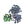
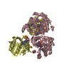
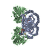
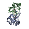
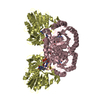
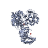
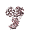
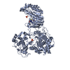
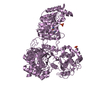
[English] 日本語
Yorodumi
- PDB-5ue9: WT DHODB with orotate bound -

+
Open data


ID or keywords:

Loading...

-
Basic information

Entry
Database: PDB / ID: 5ue9
TitleWT DHODB with orotate bound
Components(Dihydroorotate ...) x 2
KeywordsOXIDOREDUCTASE / orotate / pyrimidine biosynthesis / FeS cluster / electron transfer / dehydrogenase / protein engineering
Function / homology
Function and homology information


dihydroorotate dehydrogenase (NAD+) / dihydroorotate dehydrogenase (NAD+) activity / 'de novo' UMP biosynthetic process / 'de novo' pyrimidine nucleobase biosynthetic process / 2 iron, 2 sulfur cluster binding / flavin adenine dinucleotide binding / electron transfer activity / metal ion binding / cytoplasm
Similarity search - Function
Dihydroorotate dehydrogenase B (pyrk subunit); domain 3 / Dihydroorotate dehydrogenase, electron transfer subunit / Dihydroorotate dehydrogenase electron transfer subunit / Dihydroorotate dehydrogenase, class 1B / Dihydroorotate dehydrogenase, electron transfer subunit, Fe-S binding domain superfamily / Cytochrome-c3 hydrogenase, gamma subunit / Dihydroorotate dehydrogenase, electron transfer subunit, iron-sulphur cluster binding domain / Iron-sulfur cluster binding domain of dihydroorotate dehydrogenase B / Dihydroorotate dehydrogenase, class 1 / Dihydroorotate dehydrogenase, class 1/ 2 ...Dihydroorotate dehydrogenase B (pyrk subunit); domain 3 / Dihydroorotate dehydrogenase, electron transfer subunit / Dihydroorotate dehydrogenase electron transfer subunit / Dihydroorotate dehydrogenase, class 1B / Dihydroorotate dehydrogenase, electron transfer subunit, Fe-S binding domain superfamily / Cytochrome-c3 hydrogenase, gamma subunit / Dihydroorotate dehydrogenase, electron transfer subunit, iron-sulphur cluster binding domain / Iron-sulfur cluster binding domain of dihydroorotate dehydrogenase B / Dihydroorotate dehydrogenase, class 1 / Dihydroorotate dehydrogenase, class 1/ 2 / Dihydroorotate dehydrogenase signature 1. / Dihydroorotate dehydrogenase signature 2. / Dihydroorotate dehydrogenase, conserved site / Nucleotide-binding domain of ferredoxin-NADP reductase (FNR) module / Dihydroorotate dehydrogenase domain / Dihydroorotate dehydrogenase / Translation factors / Elongation Factor Tu (Ef-tu); domain 3 / Oxidoreductase FAD/NAD(P)-binding / Oxidoreductase NAD-binding domain / FAD-binding domain, ferredoxin reductase-type / Ferredoxin-NADP reductase (FNR), nucleotide-binding domain / Ferredoxin reductase-type FAD binding domain profile. / Riboflavin synthase-like beta-barrel / Ribbon / Aldolase class I / Aldolase-type TIM barrel / TIM Barrel / Alpha-Beta Barrel / Beta Barrel / Rossmann fold / 3-Layer(aba) Sandwich / Mainly Beta / Alpha Beta
Similarity search - Domain/homology
FLAVIN-ADENINE DINUCLEOTIDE / FE2/S2 (INORGANIC) CLUSTER / FLAVIN MONONUCLEOTIDE / OROTIC ACID / : / : / Dihydroorotate dehydrogenase B (NAD(+)), electron transfer subunit / Dihydroorotate dehydrogenase B (NAD(+)), catalytic subunit
Similarity search - Component
Biological speciesLactococcus lactis subsp. lactis (lactic acid bacteria)
MethodX-RAY DIFFRACTION / MOLECULAR REPLACEMENT / molecular replacement / Resolution: 2.72 Å
AuthorsPompeu, Y.A. / Stewart, J.D.
CitationJournal: to be published
Title: Isoleucine 74 Plays a Key Role in Controlling Electron Transfer Between Lactococcus lactis Dihydroorotate Dehydrogenase 1B Subunits
Authors: Pompeu, Y.A. / Stewart, J.D.
History
DepositionDec 29, 2016Deposition site: RCSB / Processing site: RCSB
Revision 1.0Sep 6, 2017Provider: repository / Type: Initial release
Revision 1.1Oct 4, 2023Group: Data collection / Database references / Refinement description
Category: chem_comp_atom / chem_comp_bond ...chem_comp_atom / chem_comp_bond / database_2 / pdbx_initial_refinement_model / struct_ncs_dom_lim
Item: _database_2.pdbx_DOI / _database_2.pdbx_database_accession ..._database_2.pdbx_DOI / _database_2.pdbx_database_accession / _struct_ncs_dom_lim.beg_auth_comp_id / _struct_ncs_dom_lim.beg_label_asym_id / _struct_ncs_dom_lim.beg_label_comp_id / _struct_ncs_dom_lim.beg_label_seq_id / _struct_ncs_dom_lim.end_auth_comp_id / _struct_ncs_dom_lim.end_label_asym_id / _struct_ncs_dom_lim.end_label_comp_id / _struct_ncs_dom_lim.end_label_seq_id

-
Structure visualization

Structure viewerMolecule:
MolmilJmol/JSmol

Downloads & links

-
Assembly

Deposited unit
A: Dihydroorotate dehydrogenase
B: Dihydroorotate dehydrogenase B (NAD(+)), electron transfer subunit
D: Dihydroorotate dehydrogenase B (NAD(+)), electron transfer subunit
C: Dihydroorotate dehydrogenase
hetero molecules


Theoretical massNumber of molelcules
Total (without water)125,77815
Polymers122,4104
Non-polymers3,36711
Water3,585199
1
A: Dihydroorotate dehydrogenase
B: Dihydroorotate dehydrogenase B (NAD(+)), electron transfer subunit
hetero molecules

A: Dihydroorotate dehydrogenase
B: Dihydroorotate dehydrogenase B (NAD(+)), electron transfer subunit
hetero molecules


Theoretical massNumber of molelcules
Total (without water)125,81316
Polymers122,4104
Non-polymers3,40312
Water724
TypeNameSymmetry operationNumber
identity operation1_555x,y,z1
crystal symmetry operation4_555x,-y,-z1
Buried area15560 Å2
ΔGint-126 kcal/mol
Surface area40590 Å2
MethodPISA
2
D: Dihydroorotate dehydrogenase B (NAD(+)), electron transfer subunit
C: Dihydroorotate dehydrogenase
hetero molecules

D: Dihydroorotate dehydrogenase B (NAD(+)), electron transfer subunit
C: Dihydroorotate dehydrogenase
hetero molecules


Theoretical massNumber of molelcules
Total (without water)125,74214
Polymers122,4104
Non-polymers3,33210
Water724
TypeNameSymmetry operationNumber
identity operation1_555x,y,z1
crystal symmetry operation3_454-x-1,y,-z-1/21
Buried area15680 Å2
ΔGint-114 kcal/mol
Surface area39880 Å2
MethodPISA
Unit cell
Length a, b, c (Å)80.207, 151.911, 214.799
Angle α, β, γ (deg.)90.000, 90.000, 90.000
Int Tables number20
Space group name H-MC2221
Noncrystallographic symmetry (NCS)NCS domain:
IDEns-IDDetails
11(chain A and (resid 5 through 134 or resid 136 through 309))
21(chain C and (resid 5 through 134 or resid 136 through 309))
12(chain B and (resid 1 through 9 or (resid 10...
22chain D

NCS domain segments:
Dom-IDComponent-IDEns-IDBeg auth comp-IDBeg label comp-IDEnd auth comp-IDEnd label comp-IDSelection detailsAuth asym-IDLabel asym-IDAuth seq-IDLabel seq-ID
111ASNASNSERSER(chain A and (resid 5 through 134 or resid 136 through 309))AA5 - 1341 - 130
121PROPROSERSER(chain A and (resid 5 through 134 or resid 136 through 309))AA136 - 309132 - 305
211ASNASNSERSER(chain C and (resid 5 through 134 or resid 136 through 309))CD5 - 1341 - 130
221PROPROSERSER(chain C and (resid 5 through 134 or resid 136 through 309))CD136 - 309132 - 305
112METMETTHRTHR(chain B and (resid 1 through 9 or (resid 10...BB1 - 91 - 9
122ILEILEVALVAL(chain B and (resid 1 through 9 or (resid 10...BB10 - 1110 - 11
132METMETLEULEU(chain B and (resid 1 through 9 or (resid 10...BB1 - 2621 - 262
142METMETLEULEU(chain B and (resid 1 through 9 or (resid 10...BB1 - 2621 - 262
152METMETLEULEU(chain B and (resid 1 through 9 or (resid 10...BB1 - 2621 - 262
162METMETLEULEU(chain B and (resid 1 through 9 or (resid 10...BB1 - 2621 - 262
172METMETLEULEU(chain B and (resid 1 through 9 or (resid 10...BB1 - 2621 - 262
182METMETLEULEU(chain B and (resid 1 through 9 or (resid 10...BB1 - 2621 - 262
212METMETLEULEUchain DDC1 - 2621 - 262

NCS ensembles :
ID
1
2

-
Components

-
Dihydroorotate ... , 2 types, 4 molecules ACBD

#1: Protein Dihydroorotate dehydrogenase / / DHOdehase


Mass: 32342.562 Da / Num. of mol.: 2
Source method: isolated from a genetically manipulated source
Source: (gene. exp.) Lactococcus lactis subsp. lactis (lactic acid bacteria)
Gene: pyrD, NCDO895_2365 / Plasmid: pET-22(b)+ / Production host: Escherichia coli (E. coli)
References: UniProt: A0A0V8EUT4, UniProt: Q9CFW8*PLUS, dihydroorotate dehydrogenase (NAD+)
#2: Protein Dihydroorotate dehydrogenase B (NAD(+)), electron transfer subunit / Dihydroorotate oxidase B / electron transfer subunit


Mass: 28862.615 Da / Num. of mol.: 2
Source method: isolated from a genetically manipulated source
Source: (gene. exp.) Lactococcus lactis subsp. lactis (lactic acid bacteria)
Gene: pyrK, NCDO895_2366 / Plasmid: pet 22b+ / Production host: Escherichia coli (E. coli) / References: UniProt: A0A0V8EGX5, UniProt: Q9CFW7*PLUS

-
Non-polymers , 7 types, 210 molecules

#3: Chemical ChemComp-FMN / FLAVIN MONONUCLEOTIDE / RIBOFLAVIN MONOPHOSPHATE / Flavin mononucleotide


Mass: 456.344 Da / Num. of mol.: 2 / Source method: obtained synthetically / Formula: C17H21N4O9P
#4: Chemical ChemComp-ORO / OROTIC ACID / Orotic acid


Mass: 156.096 Da / Num. of mol.: 2 / Source method: obtained synthetically / Formula: C5H4N2O4
#5: Chemical ChemComp-CL / CHLORIDE ION / Chloride


Mass: 35.453 Da / Num. of mol.: 1 / Source method: obtained synthetically / Formula: Cl
#6: Chemical ChemComp-FAD / FLAVIN-ADENINE DINUCLEOTIDE / Flavin adenine dinucleotide


Mass: 785.550 Da / Num. of mol.: 2 / Source method: obtained synthetically / Formula: C27H33N9O15P2 / Comment: FAD*YM
#7: Chemical ChemComp-FES / FE2/S2 (INORGANIC) CLUSTER / Iron–sulfur cluster


Mass: 175.820 Da / Num. of mol.: 2 / Source method: obtained synthetically / Formula: Fe2S2
#8: Chemical ChemComp-GOL / GLYCEROL / GLYCERIN / PROPANE-1,2,3-TRIOL / Glycerol


Mass: 92.094 Da / Num. of mol.: 2 / Source method: obtained synthetically / Formula: C3H8O3
#9: Water ChemComp-HOH / water / Water


Mass: 18.015 Da / Num. of mol.: 199 / Source method: isolated from a natural source / Formula: H2O

-
Experimental details

-
Experiment

ExperimentMethod: X-RAY DIFFRACTION / Number of used crystals: 1

-
Sample preparation

CrystalDensity Matthews: 2.67 Å3/Da / Density % sol: 53.98 %
Crystal growTemperature: 298 K / Method: vapor diffusion, hanging drop / pH: 4
Details: 0.1M NaAcetate pH 4, PEG 4k 12% w/v, glycerol 3% v/v

-
Data collection

DiffractionMean temperature: 298 K
Diffraction sourceSource: ROTATING ANODE / Type: RIGAKU RU200 / Wavelength: 1.54 Å
DetectorType: RIGAKU RAXIS IV++ / Detector: IMAGE PLATE / Date: Aug 10, 2012
RadiationProtocol: SINGLE WAVELENGTH / Monochromatic (M) / Laue (L): M / Scattering type: x-ray
Radiation wavelengthWavelength: 1.54 Å / Relative weight: 1
ReflectionResolution: 2.72→53 Å / Num. obs: 34552 / % possible obs: 99 % / Observed criterion σ(I): 2 / Redundancy: 5.1 % / Biso Wilson estimate: 50.8 Å2 / Rsym value: 0.09 / Net I/σ(I): 7.34

-
Phasing

PhasingMethod: molecular replacement

-
Processing

Software
NameVersionClassification
HKL-2000data collection
SCALEPACKdata scaling
PHASERphasing
PHENIXdev_2621refinement
PDB_EXTRACT3.22data extraction
iMOSFLMdata reduction
HKL-2000data reduction
RefinementMethod to determine structure: MOLECULAR REPLACEMENT
Starting model: 1EP2
Resolution: 2.72→50.389 Å / SU ML: 0.42 / Cross valid method: FREE R-VALUE / σ(F): 1.33 / Phase error: 27.04
RfactorNum. reflection% reflection
Rfree0.2543 1731 5.03 %
Rwork0.1949 --
obs0.198 34435 96.5 %
Solvent computationShrinkage radii: 0.9 Å / VDW probe radii: 1.11 Å
Displacement parametersBiso max: 105.43 Å2 / Biso mean: 42.4345 Å2 / Biso min: 17.57 Å2
Refinement stepCycle: final / Resolution: 2.72→50.389 Å
ProteinNucleic acidLigandSolventTotal
Num. atoms8541 0 211 199 8951
Biso mean--37.28 37.14 -
Num. residues----1135
Refine LS restraints
Refine-IDTypeDev idealNumber
X-RAY DIFFRACTIONf_bond_d0.0098913
X-RAY DIFFRACTIONf_angle_d1.21412100
X-RAY DIFFRACTIONf_chiral_restr0.0641400
X-RAY DIFFRACTIONf_plane_restr0.0081530
X-RAY DIFFRACTIONf_dihedral_angle_d13.515411
Refine LS restraints NCS
Ens-IDDom-IDAuth asym-IDNumberRefine-IDRmsType
11A2848X-RAY DIFFRACTION10.555TORSIONAL
12C2848X-RAY DIFFRACTION10.555TORSIONAL
21B2493X-RAY DIFFRACTION10.555TORSIONAL
22D2493X-RAY DIFFRACTION10.555TORSIONAL
LS refinement shell

Refine-ID: X-RAY DIFFRACTION / Rfactor Rfree error: 0 / Total num. of bins used: 12

Resolution (Å)Rfactor RfreeNum. reflection RfreeRfactor RworkNum. reflection RworkNum. reflection all% reflection obs (%)
2.7201-2.80010.3911440.29022772291699
2.8001-2.89050.32871460.2562767291399
2.8905-2.99380.29211560.2552729288599
2.9938-3.11370.31711240.23792747287198
3.1137-3.25530.33051470.2322708285597
3.2553-3.42690.3151400.23012713285396
3.4269-3.64160.30981500.2032691284196
3.6416-3.92270.25731440.1922721286597
3.9227-4.31720.2031420.16952711285396
4.3172-4.94150.1911460.14292679282595
4.9415-6.22390.23321370.17552697283494
6.2239-50.3980.20261550.17632769292493

+
About Yorodumi

-
News

-
Feb 9, 2022. New format data for meta-information of EMDB entries

New format data for meta-information of EMDB entries

  • Version 3 of the EMDB header file is now the official format.
  • The previous official version 1.9 will be removed from the archive.

Related info.:EMDB header

External links:wwPDB to switch to version 3 of the EMDB data model

-
Aug 12, 2020. Covid-19 info

Covid-19 info

URL: https://pdbj.org/emnavi/covid19.php

New page: Covid-19 featured information page in EM Navigator.

Related info.:Covid-19 info / Mar 5, 2020. Novel coronavirus structure data

+
Mar 5, 2020. Novel coronavirus structure data

Novel coronavirus structure data

Related info.:Yorodumi Speices / Aug 12, 2020. Covid-19 info

External links:COVID-19 featured content - PDBj / Molecule of the Month (242):Coronavirus Proteases

+
Jan 31, 2019. EMDB accession codes are about to change! (news from PDBe EMDB page)

EMDB accession codes are about to change! (news from PDBe EMDB page)

  • The allocation of 4 digits for EMDB accession codes will soon come to an end. Whilst these codes will remain in use, new EMDB accession codes will include an additional digit and will expand incrementally as the available range of codes is exhausted. The current 4-digit format prefixed with “EMD-” (i.e. EMD-XXXX) will advance to a 5-digit format (i.e. EMD-XXXXX), and so on. It is currently estimated that the 4-digit codes will be depleted around Spring 2019, at which point the 5-digit format will come into force.
  • The EM Navigator/Yorodumi systems omit the EMD- prefix.

Related info.:Q: What is EMD? / ID/Accession-code notation in Yorodumi/EM Navigator

External links:EMDB Accession Codes are Changing Soon! / Contact to PDBj

+
Jul 12, 2017. Major update of PDB

Major update of PDB

  • wwPDB released updated PDB data conforming to the new PDBx/mmCIF dictionary.
  • This is a major update changing the version number from 4 to 5, and with Remediation, in which all the entries are updated.
  • In this update, many items about electron microscopy experimental information are reorganized (e.g. em_software).
  • Now, EM Navigator and Yorodumi are based on the updated data.

External links:wwPDB Remediation / Enriched Model Files Conforming to OneDep Data Standards Now Available in the PDB FTP Archive

-
Yorodumi

Thousand views of thousand structures

  • Yorodumi is a browser for structure data from EMDB, PDB, SASBDB, etc.
  • This page is also the successor to EM Navigator detail page, and also detail information page/front-end page for Omokage search.
  • The word "yorodu" (or yorozu) is an old Japanese word meaning "ten thousand". "mi" (miru) is to see.

Related info.:EMDB / PDB / SASBDB / Comparison of 3 databanks / Yorodumi Search / Aug 31, 2016. New EM Navigator & Yorodumi / Yorodumi Papers / Jmol/JSmol / Function and homology information / Changes in new EM Navigator and Yorodumi

Read more