 Movie
Movie Controller


+ Open data

- Basic information
| Entry | Database: PDB / ID: 6czf | ||||||
|---|---|---|---|---|---|---|---|
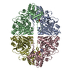
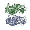
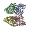
| Title | The structure of E. coli PurF in complex with ppGpp-Mg | ||||||
 Components Components | Amidophosphoribosyltransferase | ||||||
 Keywords Keywords | TRANSFERASE / PurF / ppGpp | ||||||
| Function / homology |  Function and homology informationamidophosphoribosyltransferase / amidophosphoribosyltransferase activity / purine nucleobase biosynthetic process / purine nucleotide biosynthetic process / 'de novo' IMP biosynthetic process / guanosine tetraphosphate binding / glutamine metabolic process / glycosyltransferase activity / magnesium ion binding / identical protein binding ...amidophosphoribosyltransferase / amidophosphoribosyltransferase activity / purine nucleobase biosynthetic process / purine nucleotide biosynthetic process / 'de novo' IMP biosynthetic process / guanosine tetraphosphate binding / glutamine metabolic process / glycosyltransferase activity / magnesium ion binding / identical protein binding / cytosol / cytoplasm Function and homology informationamidophosphoribosyltransferase / amidophosphoribosyltransferase activity / purine nucleobase biosynthetic process / purine nucleotide biosynthetic process / 'de novo' IMP biosynthetic process / guanosine tetraphosphate binding / glutamine metabolic process / glycosyltransferase activity / magnesium ion binding / identical protein binding ...amidophosphoribosyltransferase / amidophosphoribosyltransferase activity / purine nucleobase biosynthetic process / purine nucleotide biosynthetic process / 'de novo' IMP biosynthetic process / guanosine tetraphosphate binding / glutamine metabolic process / glycosyltransferase activity / magnesium ion binding / identical protein binding / cytosol / cytoplasmSimilarity search - Function | ||||||
| Biological species |  Escherichia coli (E. coli) Escherichia coli (E. coli) | ||||||
| Method | X-RAY DIFFRACTION / SYNCHROTRON / MOLECULAR REPLACEMENT / molecular replacement / Resolution: 1.949 Å | ||||||
 Authors Authors | Wang, B. / Grant, R.A. / Laub, M.T. | ||||||
| Funding support |  United States, 1items United States, 1items
| ||||||
 Citation Citation | Journal: Nat. Chem. Biol. / Year: 2019 Title: Affinity-based capture and identification of protein effectors of the growth regulator ppGpp. Authors: Wang, B. / Dai, P. / Ding, D. / Del Rosario, A. / Grant, R.A. / Pentelute, B.L. / Laub, M.T. | ||||||
| History |
|
- Structure visualization
| Structure viewer | Molecule: MolmilJmol/JSmol |
|---|
- Downloads & links
-Download
| PDBx/mmCIF format | 6czf.cif.gz | 729.5 KB | Display | PDBx/mmCIF format |
|---|---|---|---|---|
| PDB format | pdb6czf.ent.gz | 609.6 KB | Display | PDB format |
| PDBx/mmJSON format | 6czf.json.gz | Tree view | PDBx/mmJSON format | |
| Others |  Other downloads Other downloads |
-Validation report
| Arichive directory | https://data.pdbj.org/pub/pdb/validation_reports/cz/6czfftp://data.pdbj.org/pub/pdb/validation_reports/cz/6czf | HTTPS FTP |
|---|
-Related structure data
| Related structure data |  1ecjS S: Starting model for refinement |
|---|---|
| Similar structure data |


-Links
 PDBj
PDBj


- Assembly
| Deposited unit | 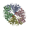
| ||||||||
|---|---|---|---|---|---|---|---|---|---|
| 1 |
| ||||||||
| Unit cell |
|
-Components
| #1: Protein | / ATase / Glutamine phosphoribosylpyrophosphate amidotransferase / GPATase Mass: 55723.961 Da / Num. of mol.: 4 Source method: isolated from a genetically manipulated source Source: (gene. exp.) Escherichia coli (strain K12) (bacteria)Strain: K12 / Gene: purF, b2312, JW2309 / Production host: Escherichia coli (E. coli) / References: UniProt: P0AG16, amidophosphoribosyltransferase#2: Chemical |   Mass: 24.305 Da / Num. of mol.: 2 / Source method: obtained synthetically / Formula: Mg Mass: 24.305 Da / Num. of mol.: 2 / Source method: obtained synthetically / Formula: Mg#3: Chemical | Guanosine pentaphosphate  Type: RNA linking / Mass: 603.160 Da / Num. of mol.: 2 / Source method: obtained synthetically / Formula: C10H17N5O17P4 / Feature type: SUBJECT OF INVESTIGATION Type: RNA linking / Mass: 603.160 Da / Num. of mol.: 2 / Source method: obtained synthetically / Formula: C10H17N5O17P4 / Feature type: SUBJECT OF INVESTIGATION#4: Water | ChemComp-HOH / | Water Mass: 18.015 Da / Num. of mol.: 380 / Source method: isolated from a natural source / Formula: H2O Mass: 18.015 Da / Num. of mol.: 380 / Source method: isolated from a natural source / Formula: H2O |
|---|
-Experimental details
-Experiment
| Experiment | Method: X-RAY DIFFRACTION / Number of used crystals: 1 |
|---|
- Sample preparation
| Crystal | Density Matthews: 2.17 Å3/Da / Density % sol: 43.22 % |
|---|---|
| Crystal grow | Temperature: 291 K / Method: vapor diffusion, hanging drop Details: Drops containing 2 uL of protein (25 mg.ml in 20 mM HEPES pH 7.4, 150 mM NaCl, 1 mM TCEP, 5 mM ppGpp, 15 mM MgCl2) were mixed with 2 uL of well solution (0.1 M HEPES pH 7.4, 24% PEG-3350, 4% isopropanol.) |
-Data collection
| Diffraction | Mean temperature: 100 K | |||||||||||||||||||||||||||||||||||||||||||||||||||||||||||||||||||||||||||||||||||||||||||||||||||||||||||||||||||||||||||||||||||||||||||||||||||||||||||||||||||||||||||||||||||||||||||||
|---|---|---|---|---|---|---|---|---|---|---|---|---|---|---|---|---|---|---|---|---|---|---|---|---|---|---|---|---|---|---|---|---|---|---|---|---|---|---|---|---|---|---|---|---|---|---|---|---|---|---|---|---|---|---|---|---|---|---|---|---|---|---|---|---|---|---|---|---|---|---|---|---|---|---|---|---|---|---|---|---|---|---|---|---|---|---|---|---|---|---|---|---|---|---|---|---|---|---|---|---|---|---|---|---|---|---|---|---|---|---|---|---|---|---|---|---|---|---|---|---|---|---|---|---|---|---|---|---|---|---|---|---|---|---|---|---|---|---|---|---|---|---|---|---|---|---|---|---|---|---|---|---|---|---|---|---|---|---|---|---|---|---|---|---|---|---|---|---|---|---|---|---|---|---|---|---|---|---|---|---|---|---|---|---|---|---|---|---|---|---|
| Diffraction source | Source: SYNCHROTRON / Site: APS / Beamline: 24-ID-C / Wavelength: 0.9791 Å | |||||||||||||||||||||||||||||||||||||||||||||||||||||||||||||||||||||||||||||||||||||||||||||||||||||||||||||||||||||||||||||||||||||||||||||||||||||||||||||||||||||||||||||||||||||||||||||
| Detector | Type: DECTRIS PILATUS3 S 6M / Detector: PIXEL / Date: Oct 1, 2017 | |||||||||||||||||||||||||||||||||||||||||||||||||||||||||||||||||||||||||||||||||||||||||||||||||||||||||||||||||||||||||||||||||||||||||||||||||||||||||||||||||||||||||||||||||||||||||||||
| Radiation | Protocol: SINGLE WAVELENGTH / Monochromatic (M) / Laue (L): M / Scattering type: x-ray | |||||||||||||||||||||||||||||||||||||||||||||||||||||||||||||||||||||||||||||||||||||||||||||||||||||||||||||||||||||||||||||||||||||||||||||||||||||||||||||||||||||||||||||||||||||||||||||
| Radiation wavelength | Wavelength: 0.9791 Å / Relative weight: 1 | |||||||||||||||||||||||||||||||||||||||||||||||||||||||||||||||||||||||||||||||||||||||||||||||||||||||||||||||||||||||||||||||||||||||||||||||||||||||||||||||||||||||||||||||||||||||||||||
| Reflection | Resolution: 1.949→100 Å / Num. obs: 141205 / % possible obs: 99.9 % / Redundancy: 6.4 % / Biso Wilson estimate: 41.33 Å2 / Rmerge(I) obs: 0.087 / Rpim(I) all: 0.037 / Rrim(I) all: 0.094 / Χ2: 0.937 / Net I/σ(I): 4.8 | |||||||||||||||||||||||||||||||||||||||||||||||||||||||||||||||||||||||||||||||||||||||||||||||||||||||||||||||||||||||||||||||||||||||||||||||||||||||||||||||||||||||||||||||||||||||||||||
| Reflection shell | Diffraction-ID: 1
|
-Phasing
| Phasing | Method: molecular replacement |
|---|
- Processing
| Software |
| |||||||||||||||||||||||||||||||||||||||||||||||||||||||||||||||||||||||||||||||||||||||||||||||||||||||||
|---|---|---|---|---|---|---|---|---|---|---|---|---|---|---|---|---|---|---|---|---|---|---|---|---|---|---|---|---|---|---|---|---|---|---|---|---|---|---|---|---|---|---|---|---|---|---|---|---|---|---|---|---|---|---|---|---|---|---|---|---|---|---|---|---|---|---|---|---|---|---|---|---|---|---|---|---|---|---|---|---|---|---|---|---|---|---|---|---|---|---|---|---|---|---|---|---|---|---|---|---|---|---|---|---|---|---|
| Refinement | Method to determine structure: MOLECULAR REPLACEMENT Starting model: 1ecj Resolution: 1.949→48.642 Å / SU ML: 0.32 / Cross valid method: THROUGHOUT / σ(F): 1.34 / Phase error: 32.85
| |||||||||||||||||||||||||||||||||||||||||||||||||||||||||||||||||||||||||||||||||||||||||||||||||||||||||
| Solvent computation | Shrinkage radii: 0.9 Å / VDW probe radii: 1.11 Å | |||||||||||||||||||||||||||||||||||||||||||||||||||||||||||||||||||||||||||||||||||||||||||||||||||||||||
| Displacement parameters | Biso max: 120.42 Å2 / Biso mean: 52.1422 Å2 / Biso min: 23.34 Å2 | |||||||||||||||||||||||||||||||||||||||||||||||||||||||||||||||||||||||||||||||||||||||||||||||||||||||||
| Refinement step | Cycle: final / Resolution: 1.949→48.642 Å
| |||||||||||||||||||||||||||||||||||||||||||||||||||||||||||||||||||||||||||||||||||||||||||||||||||||||||
| LS refinement shell | Refine-ID: X-RAY DIFFRACTION / Rfactor Rfree error: 0 / Total num. of bins used: 14
|
