 Movie
Movie Controller


+ Open data

- Basic information
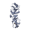
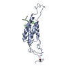
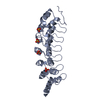
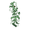
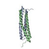
| Entry | Database: PDB / ID: 6csv | ||||||
|---|---|---|---|---|---|---|---|
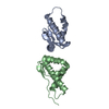
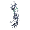
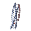
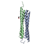
| Title | The structure of the Cep63-Cep152 heterotetrameric complex | ||||||
 Components Components | Centrosomal protein of 63 kDa,Centrosomal protein of 152 kDa Centrosome Centrosome | ||||||
 Keywords Keywords | CELL CYCLE / Centrosome / Complex / Hydrophobic | ||||||
| Function / homology |  Function and homology information Function and homology informationnegative regulation of protein K63-linked ubiquitination / de novo centriole assembly involved in multi-ciliated epithelial cell differentiation / procentriole / deuterosome / procentriole replication complex / cell projection organization / pericentriolar material / centriole replication / centrosome duplication / centriolar satellite ...negative regulation of protein K63-linked ubiquitination / de novo centriole assembly involved in multi-ciliated epithelial cell differentiation / procentriole / deuterosome / procentriole replication complex / cell projection organization / pericentriolar material / centriole replication / centrosome duplication / centriolar satellite / spindle assembly / signal transduction in response to DNA damage / Loss of Nlp from mitotic centrosomes / Loss of proteins required for interphase microtubule organization from the centrosome / Recruitment of mitotic centrosome proteins and complexes / Recruitment of NuMA to mitotic centrosomes / Anchoring of the basal body to the plasma membrane / centriole / negative regulation of innate immune response / AURKA Activation by TPX2 / DNA damage checkpoint signaling / spindle pole / Regulation of PLK1 Activity at G2/M Transition / protein stabilization / molecular adaptor activity / cell division / centrosome / protein kinase binding / nucleoplasm / cytosolSimilarity search - Function | ||||||
| Biological species |  Homo sapiens (human) Homo sapiens (human) | ||||||
| Method | X-RAY DIFFRACTION / SYNCHROTRON / SAD / Resolution: 2.5 Å | ||||||
 Authors Authors | Lee, E. / Chen, Y. / Zhang, L. / Kim, T.S. / Ahn, J.I. / Park, J.E. / Lee, K.S. | ||||||
 Citation Citation | Journal: Nat Commun / Year: 2019 Title: Molecular architecture of a cylindrical self-assembly at human centrosomes. Authors: Kim, T.S. / Zhang, L. / Il Ahn, J. / Meng, L. / Chen, Y. / Lee, E. / Bang, J.K. / Lim, J.M. / Ghirlando, R. / Fan, L. / Wang, Y.X. / Kim, B.Y. / Park, J.E. / Lee, K.S. | ||||||
| History |
|
- Structure visualization
| Structure viewer | Molecule: MolmilJmol/JSmol |
|---|
- Downloads & links
-Download
| PDBx/mmCIF format | 6csv.cif.gz | 83.4 KB | Display | PDBx/mmCIF format |
|---|---|---|---|---|
| PDB format | pdb6csv.ent.gz | 64.6 KB | Display | PDB format |
| PDBx/mmJSON format | 6csv.json.gz | Tree view | PDBx/mmJSON format | |
| Others |  Other downloads Other downloads |
-Validation report
| Arichive directory | https://data.pdbj.org/pub/pdb/validation_reports/cs/6csvftp://data.pdbj.org/pub/pdb/validation_reports/cs/6csv | HTTPS FTP |
|---|
-Related structure data


-Links
 PDBj
PDBj- Assembly
| Deposited unit | 
| ||||||||
|---|---|---|---|---|---|---|---|---|---|
| 1 | 
| ||||||||
| 2 | 
| ||||||||
| Unit cell |
|
-Components
| #1: Protein | Centrosome / Cep63 / Cep152 Mass: 11088.716 Da / Num. of mol.: 4 Source method: isolated from a genetically manipulated source Source: (gene. exp.) Homo sapiens (human) / Gene: CEP63, CEP152, KIAA0912 / Production host:  Escherichia coli (E. coli) / References: UniProt: Q96MT8, UniProt: O94986 Escherichia coli (E. coli) / References: UniProt: Q96MT8, UniProt: O94986#2: Water | ChemComp-HOH / | Water Mass: 18.015 Da / Num. of mol.: 37 / Source method: isolated from a natural source / Formula: H2O Mass: 18.015 Da / Num. of mol.: 37 / Source method: isolated from a natural source / Formula: H2O |
|---|
-Experimental details
-Experiment
| Experiment | Method: X-RAY DIFFRACTION / Number of used crystals: 1 |
|---|
- Sample preparation
| Crystal | Density Matthews: 2.51 Å3/Da / Density % sol: 51.03 % |
|---|---|
| Crystal grow | Temperature: 293 K / Method: vapor diffusion, hanging drop / Details: 0.1 M Bis-Tris (pH 5.5) and 10% PEG 3350 |
-Data collection
| Diffraction | Mean temperature: 100 K |
|---|---|
| Diffraction source | Source: SYNCHROTRON / Site: APS  / Beamline: 22-ID / Wavelength: 0.9793 Å / Beamline: 22-ID / Wavelength: 0.9793 Å |
| Detector | Type: DECTRIS PILATUS3 R CdTe 300K-W / Detector: PIXEL / Date: Mar 4, 2016 |
| Radiation | Protocol: SINGLE WAVELENGTH / Monochromatic (M) / Laue (L): M / Scattering type: x-ray |
| Radiation wavelength | Wavelength: 0.9793 Å / Relative weight: 1 |
| Reflection | Resolution: 2.5→19.93 Å / Num. obs: 19496 / % possible obs: 93.17 % / Redundancy: 4.9 % / Net I/σ(I): 15.82 |
| Reflection shell | Resolution: 2.5→2.54 Å |
- Processing
| Software |
| ||||||||||||||||||||||||||||||||||||||||||||||||||||||||||||||||||||||||||||||||||||||||||||||||||||||||||||||||||||||||||||||||||||||||||||||||||||||||||||||||||||||||||||||||||||||
|---|---|---|---|---|---|---|---|---|---|---|---|---|---|---|---|---|---|---|---|---|---|---|---|---|---|---|---|---|---|---|---|---|---|---|---|---|---|---|---|---|---|---|---|---|---|---|---|---|---|---|---|---|---|---|---|---|---|---|---|---|---|---|---|---|---|---|---|---|---|---|---|---|---|---|---|---|---|---|---|---|---|---|---|---|---|---|---|---|---|---|---|---|---|---|---|---|---|---|---|---|---|---|---|---|---|---|---|---|---|---|---|---|---|---|---|---|---|---|---|---|---|---|---|---|---|---|---|---|---|---|---|---|---|---|---|---|---|---|---|---|---|---|---|---|---|---|---|---|---|---|---|---|---|---|---|---|---|---|---|---|---|---|---|---|---|---|---|---|---|---|---|---|---|---|---|---|---|---|---|---|---|---|---|
| Refinement | Method to determine structure: SAD / Resolution: 2.5→19.926 Å / Cor.coef. Fo:Fc: 0.904 / Cor.coef. Fo:Fc free: 0.867 / SU B: 13.934 / SU ML: 0.3 / Cross valid method: THROUGHOUT / ESU R: 0.798 / ESU R Free: 0.388 / Details: HYDROGENS HAVE BEEN ADDED IN THE RIDING POSITIONS
| ||||||||||||||||||||||||||||||||||||||||||||||||||||||||||||||||||||||||||||||||||||||||||||||||||||||||||||||||||||||||||||||||||||||||||||||||||||||||||||||||||||||||||||||||||||||
| Solvent computation | Ion probe radii: 0.8 Å / Shrinkage radii: 0.8 Å / VDW probe radii: 1.2 Å | ||||||||||||||||||||||||||||||||||||||||||||||||||||||||||||||||||||||||||||||||||||||||||||||||||||||||||||||||||||||||||||||||||||||||||||||||||||||||||||||||||||||||||||||||||||||
| Displacement parameters | Biso mean: 57.564 Å2
| ||||||||||||||||||||||||||||||||||||||||||||||||||||||||||||||||||||||||||||||||||||||||||||||||||||||||||||||||||||||||||||||||||||||||||||||||||||||||||||||||||||||||||||||||||||||
| Refinement step | Cycle: 1 / Resolution: 2.5→19.926 Å
| ||||||||||||||||||||||||||||||||||||||||||||||||||||||||||||||||||||||||||||||||||||||||||||||||||||||||||||||||||||||||||||||||||||||||||||||||||||||||||||||||||||||||||||||||||||||
| Refine LS restraints |
|