 Movie
Movie Controller


+ Open data

- Basic information
| Entry | Database: PDB / ID: 6cme | ||||||
|---|---|---|---|---|---|---|---|
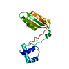
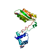
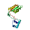
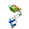
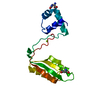
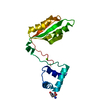
| Title | Structure of wild-type ISL2-LID in complex with LHX4-LIM1+2 | ||||||
 Components Components | LIM/homeobox protein Lhx4,Insulin gene enhancer protein ISL-2 | ||||||
 Keywords Keywords |  TRANSCRIPTION / LIM-homeodomain / cell-type specification / neural development TRANSCRIPTION / LIM-homeodomain / cell-type specification / neural development | ||||||
| Function / homology |  Function and homology information Function and homology informationvisceral motor neuron differentiation / medial motor column neuron differentiation / spinal cord motor neuron cell fate specification / neuron fate specification / neuron fate commitment / methyl-CpG binding / motor neuron axon guidance / retinal ganglion cell axon guidance / negative regulation of neuron differentiation / cis-regulatory region sequence-specific DNA binding ...visceral motor neuron differentiation / medial motor column neuron differentiation / spinal cord motor neuron cell fate specification / neuron fate specification / neuron fate commitment / methyl-CpG binding / motor neuron axon guidance / retinal ganglion cell axon guidance / negative regulation of neuron differentiation / cis-regulatory region sequence-specific DNA binding / axonogenesis / placenta development / animal organ morphogenesis / RNA polymerase II transcription regulatory region sequence-specific DNA binding / neuron differentiation / sequence-specific double-stranded DNA binding / DNA-binding transcription activator activity, RNA polymerase II-specific / sequence-specific DNA binding / DNA-binding transcription factor activity, RNA polymerase II-specific / apoptotic process / regulation of transcription by RNA polymerase II / negative regulation of apoptotic process / positive regulation of transcription by RNA polymerase II / zinc ion binding / nucleoplasm / metal ion binding / nucleusSimilarity search - Function | ||||||
| Biological species |  Mus musculus (house mouse) Mus musculus (house mouse) | ||||||
| Method | X-RAY DIFFRACTION / MOLECULAR REPLACEMENT / Resolution: 1.92 Å | ||||||
 Authors Authors | Stokes, P.H. / Silva, A. / Guss, J.M. / Matthews, J.M. | ||||||
| Funding support |  Australia, 1items Australia, 1items
| ||||||
 Citation Citation | Journal: Proteins / Year: 2019 Title: Mutation in a flexible linker modulates binding affinity for modular complexes. Authors: Philippa H Stokes / Neil O Robertson / Ana P G Silva / Tanya Estephan / Jill Trewhella / J Mitchell Guss / Jacqueline M Matthews / Abstract: Tandem beta zippers are modular complexes formed between repeated linear motifs and tandemly arrayed domains of partner proteins in which β-strands form upon binding. Studies of such complexes, ...Tandem beta zippers are modular complexes formed between repeated linear motifs and tandemly arrayed domains of partner proteins in which β-strands form upon binding. Studies of such complexes, formed by LIM domain proteins and linear motifs in their intrinsically disordered partners, revealed spacer regions between the linear motifs that are relatively flexible but may affect the overall orientation of the binding modules. We demonstrate that mutation of a solvent exposed side chain in the spacer region of an LHX4-ISL2 complex has no significant effect on the structure of the complex, but decreases binding affinity, apparently by increasing flexibility of the linker. | ||||||
| History |
|
- Structure visualization
| Structure viewer | Molecule: MolmilJmol/JSmol |
|---|
- Downloads & links
-Download
| PDBx/mmCIF format | 6cme.cif.gz | 83.1 KB | Display | PDBx/mmCIF format |
|---|---|---|---|---|
| PDB format | pdb6cme.ent.gz | 60.2 KB | Display | PDB format |
| PDBx/mmJSON format | 6cme.json.gz | Tree view | PDBx/mmJSON format | |
| Others |  Other downloads Other downloads |
-Validation report
| Arichive directory | https://data.pdbj.org/pub/pdb/validation_reports/cm/6cmeftp://data.pdbj.org/pub/pdb/validation_reports/cm/6cme | HTTPS FTP |
|---|
-Related structure data
| Related structure data |  3mmkS S: Starting model for refinement C: citing same article ( |
|---|---|
| Similar structure data | |
| Other databases |
|


-Links
 PDBj
PDBj
- Assembly
| Deposited unit | 
| ||||||||
|---|---|---|---|---|---|---|---|---|---|
| 1 | 
| ||||||||
| 2 | 
| ||||||||
| Unit cell |
| ||||||||
| Details | The authors state that light scattering and SAXS data indicate that the molecule is monomeric ion solution. SAXS data indicates a more elongated structure indicating that the extreme bend in the structure is a crystallization artifact. |
-Components
| #1: Protein | Mass: 18749.406 Da / Num. of mol.: 2 Source method: isolated from a genetically manipulated source Source: (gene. exp.) Mus musculus (house mouse) / Gene: Lhx4, Gsh-4, Gsh4, Isl2 / Details (production host): pGEX-2T / Production host:  Escherichia coli BL21(DE3) (bacteria) / Strain (production host): BL21(DE3) / References: UniProt: P53776, UniProt: Q9CXV0 Escherichia coli BL21(DE3) (bacteria) / Strain (production host): BL21(DE3) / References: UniProt: P53776, UniProt: Q9CXV0#2: Chemical | ChemComp-ZN /   Mass: 65.409 Da / Num. of mol.: 8 / Source method: obtained synthetically / Formula: Zn Mass: 65.409 Da / Num. of mol.: 8 / Source method: obtained synthetically / Formula: Zn#3: Water | ChemComp-HOH / | Water Mass: 18.015 Da / Num. of mol.: 210 / Source method: isolated from a natural source / Formula: H2O Mass: 18.015 Da / Num. of mol.: 210 / Source method: isolated from a natural source / Formula: H2O |
|---|
-Experimental details
-Experiment
| Experiment | Method: X-RAY DIFFRACTION / Number of used crystals: 1 |
|---|
- Sample preparation
| Crystal | Density Matthews: 2.47 Å3/Da / Density % sol: 53.34 % / Description: wedge-like thin sheets |
|---|---|
| Crystal grow | Temperature: 293 K / Method: vapor diffusion, sitting drop / Details: 110 mM BisTris propane and 3 M ammonium acetate |
-Data collection
| Diffraction | Mean temperature: 100 K / Serial crystal experiment: N |
|---|---|
| Diffraction source | Source: ROTATING ANODE / Type: RIGAKU MICROMAX-007 HF / Wavelength: 1.5418 Å |
| Detector | Type: MAR scanner 345 mm plate / Detector: IMAGE PLATE / Date: Nov 25, 2014 |
| Radiation | Monochromator: CONFOCAL MAXFLUX MIRRORS / Protocol: SINGLE WAVELENGTH / Monochromatic (M) / Laue (L): M / Scattering type: x-ray |
| Radiation wavelength | Wavelength: 1.5418 Å / Relative weight: 1 |
| Reflection | Resolution: 1.92→27.55 Å / Num. obs: 29762 / % possible obs: 99.8 % / Redundancy: 4.6 % / Biso Wilson estimate: 23.8 Å2 / CC1/2: 1 / Rmerge(I) obs: 0.125 / Rpim(I) all: 0.063 / Rrim(I) all: 0.14 / Net I/σ(I): 10.2 |
| Reflection shell | Resolution: 1.92→1.95 Å / Redundancy: 3.2 % / Rmerge(I) obs: 0.77 / Mean I/σ(I) obs: 1.3 / Num. unique obs: 1441 / CC1/2: 0.393 / Rpim(I) all: 0.481 / Rrim(I) all: 0.915 / % possible all: 97.6 |
- Processing
| Software |
| ||||||||||||||||||||||||||||||||||||||||||||||||||||||||||||||||||||||||||||||||||||||||||||||||||||||||||||||||||||||||||||||||||||||||||||||||||||||||||||||||||||||||||||||||||||||
|---|---|---|---|---|---|---|---|---|---|---|---|---|---|---|---|---|---|---|---|---|---|---|---|---|---|---|---|---|---|---|---|---|---|---|---|---|---|---|---|---|---|---|---|---|---|---|---|---|---|---|---|---|---|---|---|---|---|---|---|---|---|---|---|---|---|---|---|---|---|---|---|---|---|---|---|---|---|---|---|---|---|---|---|---|---|---|---|---|---|---|---|---|---|---|---|---|---|---|---|---|---|---|---|---|---|---|---|---|---|---|---|---|---|---|---|---|---|---|---|---|---|---|---|---|---|---|---|---|---|---|---|---|---|---|---|---|---|---|---|---|---|---|---|---|---|---|---|---|---|---|---|---|---|---|---|---|---|---|---|---|---|---|---|---|---|---|---|---|---|---|---|---|---|---|---|---|---|---|---|---|---|---|---|
| Refinement | Method to determine structure: MOLECULAR REPLACEMENT Starting model: 3mmk Resolution: 1.92→25 Å / Cor.coef. Fo:Fc: 0.952 / Cor.coef. Fo:Fc free: 0.929 / SU B: 4.568 / SU ML: 0.125 / Cross valid method: THROUGHOUT / ESU R: 0.154 / ESU R Free: 0.147 / Details: HYDROGENS HAVE BEEN ADDED IN THE RIDING POSITIONS
| ||||||||||||||||||||||||||||||||||||||||||||||||||||||||||||||||||||||||||||||||||||||||||||||||||||||||||||||||||||||||||||||||||||||||||||||||||||||||||||||||||||||||||||||||||||||
| Solvent computation | Ion probe radii: 0.8 Å / Shrinkage radii: 0.8 Å / VDW probe radii: 1.2 Å | ||||||||||||||||||||||||||||||||||||||||||||||||||||||||||||||||||||||||||||||||||||||||||||||||||||||||||||||||||||||||||||||||||||||||||||||||||||||||||||||||||||||||||||||||||||||
| Displacement parameters | Biso mean: 30.694 Å2
| ||||||||||||||||||||||||||||||||||||||||||||||||||||||||||||||||||||||||||||||||||||||||||||||||||||||||||||||||||||||||||||||||||||||||||||||||||||||||||||||||||||||||||||||||||||||
| Refinement step | Cycle: 1 / Resolution: 1.92→25 Å
| ||||||||||||||||||||||||||||||||||||||||||||||||||||||||||||||||||||||||||||||||||||||||||||||||||||||||||||||||||||||||||||||||||||||||||||||||||||||||||||||||||||||||||||||||||||||
| Refine LS restraints |
|