 Movie
Movie Controller

[English] 日本語
 Yorodumi
- PDB-6caf: High Resolution Structure of Concanavalin B from Jack Bean (Canav... -
+ Open data

- Basic information
| Entry | Database: PDB / ID: 6caf | ||||||
|---|---|---|---|---|---|---|---|
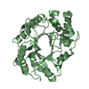
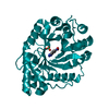
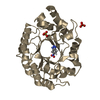
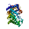
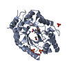
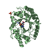
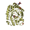
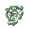
| Title | High Resolution Structure of Concanavalin B from Jack Bean (Canavalia ensiformis), A Chitinase-like Protein | ||||||
 Components Components | Concanavalin B | ||||||
 Keywords Keywords |  PLANT PROTEIN / SEED PROTEIN / chitinase-like protein / MES / Jack Bean PLANT PROTEIN / SEED PROTEIN / chitinase-like protein / MES / Jack Bean | ||||||
| Function / homology |  Function and homology information Function and homology information | ||||||
| Biological species |   Canavalia ensiformis (jack bean) Canavalia ensiformis (jack bean) | ||||||
| Method | X-RAY DIFFRACTION / SYNCHROTRON / MOLECULAR REPLACEMENT / Resolution: 1.3 Å | ||||||
 Authors Authors | McPherson, A. | ||||||
 Citation Citation | Journal: To be published Title: High Resolution Structure of Concanavalin B from Jack Bean (Canavalia ensiformis), a Chitinase-like Protein Authors: McPherson, A. | ||||||
| History |
|
- Structure visualization
| Structure viewer | Molecule: MolmilJmol/JSmol |
|---|
- Downloads & links
-Download
| PDBx/mmCIF format | 6caf.cif.gz | 146.5 KB | Display | PDBx/mmCIF format |
|---|---|---|---|---|
| PDB format | pdb6caf.ent.gz | 119.2 KB | Display | PDB format |
| PDBx/mmJSON format | 6caf.json.gz | Tree view | PDBx/mmJSON format | |
| Others |  Other downloads Other downloads |
-Validation report
| Arichive directory | https://data.pdbj.org/pub/pdb/validation_reports/ca/6cafftp://data.pdbj.org/pub/pdb/validation_reports/ca/6caf | HTTPS FTP |
|---|
-Related structure data
| Similar structure data |
|---|

-Links
 PDBj
PDBj- Assembly
| Deposited unit | 
| ||||||||
|---|---|---|---|---|---|---|---|---|---|
| 1 |
| ||||||||
| Unit cell |
|
-Components
| #1: Protein | Mass: 36762.883 Da / Num. of mol.: 1 Source method: isolated from a genetically manipulated source Source: (gene. exp.) Canavalia ensiformis (jack bean) / Production host: Canavalia ensiformis (jack bean) / References: UniProt: P49347 |
|---|---|
| #2: Water | ChemComp-HOH / Water Mass: 18.015 Da / Num. of mol.: 344 / Source method: isolated from a natural source / Formula: H2O Mass: 18.015 Da / Num. of mol.: 344 / Source method: isolated from a natural source / Formula: H2O |
-Experimental details
-Experiment
| Experiment | Method: X-RAY DIFFRACTION / Number of used crystals: 1 |
|---|
- Sample preparation
| Crystal | Density Matthews: 2.54 Å3/Da / Density % sol: 51.67 % / Description: Extremely elongated hexagonal prisms |
|---|---|
| Crystal grow | Temperature: 298 K / Method: vapor diffusion, sitting drop / pH: 6.5 Details: 2% saturated ammonium sulfate buffered with Na phosphate at pH 6.5. Room temperature, crystallization in about 1 week. Protein dissolved in 5% NaCl and 0.1 M acetic acid. PH range: 5.5 7.0 |
-Data collection
| Diffraction | Mean temperature: 173 K |
|---|---|
| Diffraction source | Source: SYNCHROTRON / Site: ALS  / Beamline: 8.3.1 / Wavelength: 0.987 Å / Beamline: 8.3.1 / Wavelength: 0.987 Å |
| Detector | Type: DECTRIS PILATUS3 R CdTe 300K-W / Detector: PIXEL / Date: Aug 20, 2016 |
| Radiation | Protocol: SINGLE WAVELENGTH / Monochromatic (M) / Laue (L): M / Scattering type: x-ray |
| Radiation wavelength | Wavelength: 0.987 Å / Relative weight: 1 |
| Reflection | Resolution: 1.3→101.31 Å / Num. obs: 73116 / % possible obs: 82 % / Redundancy: 51.5 % / CC1/2: 1 / Rmerge(I) obs: 0.167 / Rpim(I) all: 0.022 / Rrim(I) all: 0.168 / Rsym value: 0.158 / Net I/av σ(I): 50 / Net I/σ(I): 50.1 |
| Reflection shell | Resolution: 1.3→1.33 Å / Redundancy: 5.5 % / Rmerge(I) obs: 0.85 / Mean I/σ(I) obs: 3.6 / Num. unique obs: 794 / CC1/2: 0.71 / Rpim(I) all: 0.331 / Rrim(I) all: 0.918 / Rsym value: 0.78 / % possible all: 18.1 |
- Processing
| Software |
| ||||||||||||||||||||||||||||||||||||||||||||||||||||||||||||||||||||||||||||||||||||||||||||||||||||||||||||||||||||||||||||||||||||||||||||||||||||||||||||||||||||||||||||||||||||||
|---|---|---|---|---|---|---|---|---|---|---|---|---|---|---|---|---|---|---|---|---|---|---|---|---|---|---|---|---|---|---|---|---|---|---|---|---|---|---|---|---|---|---|---|---|---|---|---|---|---|---|---|---|---|---|---|---|---|---|---|---|---|---|---|---|---|---|---|---|---|---|---|---|---|---|---|---|---|---|---|---|---|---|---|---|---|---|---|---|---|---|---|---|---|---|---|---|---|---|---|---|---|---|---|---|---|---|---|---|---|---|---|---|---|---|---|---|---|---|---|---|---|---|---|---|---|---|---|---|---|---|---|---|---|---|---|---|---|---|---|---|---|---|---|---|---|---|---|---|---|---|---|---|---|---|---|---|---|---|---|---|---|---|---|---|---|---|---|---|---|---|---|---|---|---|---|---|---|---|---|---|---|---|---|
| Refinement | Method to determine structure: MOLECULAR REPLACEMENT / Resolution: 1.3→69.27 Å / Cor.coef. Fo:Fc: 0.986 / Cor.coef. Fo:Fc free: 0.978 / SU B: 0.836 / SU ML: 0.016 / Cross valid method: THROUGHOUT / ESU R: 0.034 / ESU R Free: 0.036 / Stereochemistry target values: MAXIMUM LIKELIHOOD / Details: HYDROGENS HAVE BEEN ADDED IN THE RIDING POSITIONS
| ||||||||||||||||||||||||||||||||||||||||||||||||||||||||||||||||||||||||||||||||||||||||||||||||||||||||||||||||||||||||||||||||||||||||||||||||||||||||||||||||||||||||||||||||||||||
| Solvent computation | Ion probe radii: 0.8 Å / Shrinkage radii: 0.8 Å / VDW probe radii: 1.2 Å / Solvent model: BABINET MODEL WITH MASK | ||||||||||||||||||||||||||||||||||||||||||||||||||||||||||||||||||||||||||||||||||||||||||||||||||||||||||||||||||||||||||||||||||||||||||||||||||||||||||||||||||||||||||||||||||||||
| Displacement parameters | Biso mean: 22.91 Å2
| ||||||||||||||||||||||||||||||||||||||||||||||||||||||||||||||||||||||||||||||||||||||||||||||||||||||||||||||||||||||||||||||||||||||||||||||||||||||||||||||||||||||||||||||||||||||
| Refinement step | Cycle: 1 / Resolution: 1.3→69.27 Å
| ||||||||||||||||||||||||||||||||||||||||||||||||||||||||||||||||||||||||||||||||||||||||||||||||||||||||||||||||||||||||||||||||||||||||||||||||||||||||||||||||||||||||||||||||||||||
| Refine LS restraints |
|