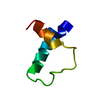
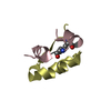
 Movie
Movie Controller


+ Open data

- Basic information
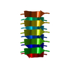
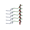
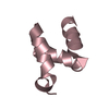
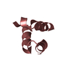
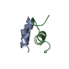
| Entry | Database: PDB / ID: 6bxx | ||||||||||||
|---|---|---|---|---|---|---|---|---|---|---|---|---|---|
| Title | GYNGFG from low-complexity domain of hnRNPA1, residues 243-248 | ||||||||||||
 Components Components | hnRNPA1 | ||||||||||||
 Keywords Keywords | PROTEIN FIBRIL / Amyloid / LARKS / Reversible-amyloid / low-complexity | ||||||||||||
| Function / homology |  Function and homology information Function and homology informationcellular response to sodium arsenite / SARS-CoV-1-host interactions / import into nucleus / telomeric repeat-containing RNA binding / G-rich strand telomeric DNA binding / pre-mRNA binding / nuclear export / RNA export from nucleus / miRNA binding / FGFR2 alternative splicing ...cellular response to sodium arsenite / SARS-CoV-1-host interactions / import into nucleus / telomeric repeat-containing RNA binding / G-rich strand telomeric DNA binding / pre-mRNA binding / nuclear export / RNA export from nucleus / miRNA binding / FGFR2 alternative splicing / regulation of alternative mRNA splicing, via spliceosome / intracellular non-membrane-bounded organelle / SARS-CoV-1 modulates host translation machinery / regulation of RNA splicing / negative regulation of telomere maintenance via telomerase / Processing of Capped Intron-Containing Pre-mRNA / mRNA transport / localization / cellular response to glucose starvation / positive regulation of telomere maintenance via telomerase / catalytic step 2 spliceosome / molecular condensate scaffold activity / mRNA Splicing - Major Pathway / mRNA 3'-UTR binding / spliceosomal complex / mRNA splicing, via spliceosome / single-stranded DNA binding / amyloid fibril formation / single-stranded RNA binding / ribonucleoprotein complex / protein domain specific binding / DNA binding / RNA binding / extracellular exosome / nucleoplasm / membrane / identical protein binding / nucleus / cytosol / cytoplasmSimilarity search - Function | ||||||||||||
| Biological species |  Homo sapiens (human) Homo sapiens (human) | ||||||||||||
| Method | X-RAY DIFFRACTION / SYNCHROTRON / AB INITIO PHASING / Resolution: 1.1 Å | ||||||||||||
 Authors Authors | Hughes, M.P. / Rodriguez, J.A. / Sawaya, M.R. / Cascio, D. / Gonen, T. / Eisenberg, D.S. | ||||||||||||
| Funding support |  United States, 3items United States, 3items
| ||||||||||||
 Citation Citation | Journal: Science / Year: 2018 Title: Atomic structures of low-complexity protein segments reveal kinked β sheets that assemble networks. Authors: Michael P Hughes / Michael R Sawaya / David R Boyer / Lukasz Goldschmidt / Jose A Rodriguez / Duilio Cascio / Lisa Chong / Tamir Gonen / David S Eisenberg / Abstract: Subcellular membraneless assemblies are a reinvigorated area of study in biology, with spirited scientific discussions on the forces between the low-complexity protein domains within these assemblies. ...Subcellular membraneless assemblies are a reinvigorated area of study in biology, with spirited scientific discussions on the forces between the low-complexity protein domains within these assemblies. To illuminate these forces, we determined the atomic structures of five segments from protein low-complexity domains associated with membraneless assemblies. Their common structural feature is the stacking of segments into kinked β sheets that pair into protofilaments. Unlike steric zippers of amyloid fibrils, the kinked sheets interact weakly through polar atoms and aromatic side chains. By computationally threading the human proteome on our kinked structures, we identified hundreds of low-complexity segments potentially capable of forming such interactions. These segments are found in proteins as diverse as RNA binders, nuclear pore proteins, and keratins, which are known to form networks and localize to membraneless assemblies. | ||||||||||||
| History |
|
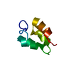
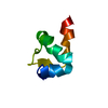
- Structure visualization
| Structure viewer | Molecule: MolmilJmol/JSmol |
|---|
- Downloads & links
-Download
| PDBx/mmCIF format | 6bxx.cif.gz | 11.3 KB | Display | PDBx/mmCIF format |
|---|---|---|---|---|
| PDB format | pdb6bxx.ent.gz | 6.3 KB | Display | PDB format |
| PDBx/mmJSON format | 6bxx.json.gz | Tree view | PDBx/mmJSON format | |
| Others |  Other downloads Other downloads |
-Validation report
| Arichive directory | https://data.pdbj.org/pub/pdb/validation_reports/bx/6bxxftp://data.pdbj.org/pub/pdb/validation_reports/bx/6bxx | HTTPS FTP |
|---|
-Related structure data


-Links
 PDBj
PDBj


- Assembly
| Deposited unit | 
| ||||||||
|---|---|---|---|---|---|---|---|---|---|
| 1 | x 10
| ||||||||
| Unit cell |
|
-Components
| #1: Protein/peptide | Mass: 613.621 Da / Num. of mol.: 1 / Fragment: residues 243-248 / Source method: obtained synthetically Details: Synthetic peptide GYNGFG corresponding tosegment 243-248 of hnRNPA1 Source: (synth.) Homo sapiens (human) / References: UniProt: P09651*PLUS |
|---|---|
| #2: Water | ChemComp-HOH / Water Mass: 18.015 Da / Num. of mol.: 4 / Source method: isolated from a natural source / Formula: H2O Mass: 18.015 Da / Num. of mol.: 4 / Source method: isolated from a natural source / Formula: H2O |
-Experimental details
-Experiment
| Experiment | Method: X-RAY DIFFRACTION / Number of used crystals: 1 |
|---|
- Sample preparation
| Crystal | Mosaicity: 0.8 ° |
|---|---|
| Crystal grow | Temperature: 298 K / Method: vapor diffusion, hanging drop / pH: 6.5 / Details: 0.1M sodium cacodylate pH 6.5, 30% (v/v) MPD |
-Data collection
| Diffraction | Mean temperature: 100 K | |||||||||||||||||||||||||||||||||||||||||||||||||||||||||||||||||||||||||||||
|---|---|---|---|---|---|---|---|---|---|---|---|---|---|---|---|---|---|---|---|---|---|---|---|---|---|---|---|---|---|---|---|---|---|---|---|---|---|---|---|---|---|---|---|---|---|---|---|---|---|---|---|---|---|---|---|---|---|---|---|---|---|---|---|---|---|---|---|---|---|---|---|---|---|---|---|---|---|---|
| Diffraction source | Source: SYNCHROTRON / Site: APS / Beamline: 24-ID-E / Wavelength: 0.9791 Å | |||||||||||||||||||||||||||||||||||||||||||||||||||||||||||||||||||||||||||||
| Detector | Type: ADSC QUANTUM 315 / Detector: CCD / Date: Mar 10, 2015 | |||||||||||||||||||||||||||||||||||||||||||||||||||||||||||||||||||||||||||||
| Radiation | Protocol: SINGLE WAVELENGTH / Monochromatic (M) / Laue (L): M / Scattering type: x-ray | |||||||||||||||||||||||||||||||||||||||||||||||||||||||||||||||||||||||||||||
| Radiation wavelength | Wavelength: 0.9791 Å / Relative weight: 1 | |||||||||||||||||||||||||||||||||||||||||||||||||||||||||||||||||||||||||||||
| Reflection | Resolution: 1.1→100 Å / Num. obs: 1269 / % possible obs: 78.9 % / Redundancy: 25.7 % / Biso Wilson estimate: 3.1 Å2 / Rmerge(I) obs: 0.13 / Χ2: 1.091 / Net I/σ(I): 8.4 / Num. measured all: 32560 | |||||||||||||||||||||||||||||||||||||||||||||||||||||||||||||||||||||||||||||
| Reflection shell |
|
- Processing
| Software |
| ||||||||||||||||||||||||
|---|---|---|---|---|---|---|---|---|---|---|---|---|---|---|---|---|---|---|---|---|---|---|---|---|---|
| Refinement | Method to determine structure: AB INITIO PHASING / Resolution: 1.1→20.486 Å / SU ML: 0.03 / Cross valid method: THROUGHOUT / σ(F): 1.56 / Phase error: 6.83
| ||||||||||||||||||||||||
| Solvent computation | Shrinkage radii: 0.9 Å / VDW probe radii: 1.11 Å | ||||||||||||||||||||||||
| Displacement parameters | Biso max: 33.87 Å2 / Biso mean: 1.9357 Å2 / Biso min: 0.53 Å2 | ||||||||||||||||||||||||
| Refinement step | Cycle: final / Resolution: 1.1→20.486 Å
| ||||||||||||||||||||||||
| Refine LS restraints |
| ||||||||||||||||||||||||
| LS refinement shell | Resolution: 1.1003→20.49 Å / Rfactor Rfree error: 0 / Total num. of bins used: 1
|
