 Movie
Movie Controller


+ Open data

- Basic information
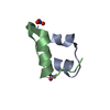
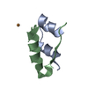
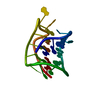
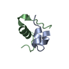
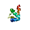
| Entry | Database: PDB / ID: 5t7r | ||||||
|---|---|---|---|---|---|---|---|
| Title | A6-A11 trans-dicarba human insulin | ||||||
 Components Components |
| ||||||
 Keywords Keywords |  HORMONE / Insulin dicarba bond HORMONE / Insulin dicarba bond | ||||||
| Function / homology |  Function and homology information Function and homology informationnegative regulation of NAD(P)H oxidase activity / negative regulation of glycogen catabolic process / regulation of cellular amino acid metabolic process / Signaling by Insulin receptor / IRS activation / negative regulation of fatty acid metabolic process / Insulin processing / negative regulation of feeding behavior / regulation of protein secretion / nitric oxide-cGMP-mediated signaling ...negative regulation of NAD(P)H oxidase activity / negative regulation of glycogen catabolic process / regulation of cellular amino acid metabolic process / Signaling by Insulin receptor / IRS activation / negative regulation of fatty acid metabolic process / Insulin processing / negative regulation of feeding behavior / regulation of protein secretion / nitric oxide-cGMP-mediated signaling / positive regulation of peptide hormone secretion / positive regulation of respiratory burst / Regulation of gene expression in beta cells / negative regulation of acute inflammatory response / alpha-beta T cell activation / negative regulation of respiratory burst involved in inflammatory response / positive regulation of dendritic spine maintenance / Synthesis, secretion, and deacylation of Ghrelin / positive regulation of glycogen biosynthetic process / negative regulation of protein secretion / Signal attenuation / FOXO-mediated transcription of oxidative stress, metabolic and neuronal genes / negative regulation of gluconeogenesis / positive regulation of nitric oxide mediated signal transduction / fatty acid homeostasis / regulation of protein localization to plasma membrane / COPI-mediated anterograde transport / negative regulation of lipid catabolic process / positive regulation of lipid biosynthetic process / negative regulation of oxidative stress-induced intrinsic apoptotic signaling pathway / positive regulation of insulin receptor signaling pathway / negative regulation of reactive oxygen species biosynthetic process / transport vesicle / positive regulation of protein autophosphorylation / Insulin receptor recycling / insulin-like growth factor receptor binding / NPAS4 regulates expression of target genes / positive regulation of protein metabolic process / positive regulation of brown fat cell differentiation / endoplasmic reticulum-Golgi intermediate compartment membrane / neuron projection maintenance / activation of protein kinase B activity / positive regulation of glycolytic process / Insulin receptor signalling cascade / positive regulation of mitotic nuclear division / positive regulation of long-term synaptic potentiation / Regulation of insulin secretion / endosome lumen / positive regulation of cytokine production / acute-phase response / positive regulation of protein secretion / positive regulation of nitric-oxide synthase activity / positive regulation of cell differentiation / regulation of transmembrane transporter activity / positive regulation of glucose import / negative regulation of proteolysis / regulation of synaptic plasticity / wound healing / insulin receptor binding / hormone activity / negative regulation of protein catabolic process / cognition / positive regulation of neuron projection development / Golgi lumen / positive regulation of protein localization to nucleus / vasodilation / glucose metabolic process / regulation of protein localization / glucose homeostasis / cell-cell signaling / insulin receptor signaling pathway / positive regulation of NF-kappaB transcription factor activity / PI5P, PP2A and IER3 Regulate PI3K/AKT Signaling / positive regulation of cell growth / secretory granule lumen / protease binding / positive regulation of MAPK cascade / positive regulation of phosphatidylinositol 3-kinase/protein kinase B signal transduction / positive regulation of cell migration / G protein-coupled receptor signaling pathway / Amyloid fiber formation / endoplasmic reticulum lumen / Golgi membrane / negative regulation of gene expression / positive regulation of cell population proliferation / positive regulation of gene expression / regulation of DNA-templated transcription / extracellular space / extracellular region / identical protein bindingSimilarity search - Function | ||||||
| Biological species |  Homo sapiens (human) Homo sapiens (human) | ||||||
| Method | X-RAY DIFFRACTION / SYNCHROTRON / Resolution: 1.55 Å | ||||||
 Authors Authors | Menting, J.G. / Lawrence, M.C. / Robinson, A.J. / Forbes, B.E. | ||||||
 Citation Citation | Journal: Sci Rep / Year: 2017 Title: Insulin in motion: The A6-A11 disulfide bond allosterically modulates structural transitions required for insulin activity. Authors: van Lierop, B. / Ong, S.C. / Belgi, A. / Delaine, C. / Andrikopoulos, S. / Haworth, N.L. / Menting, J.G. / Lawrence, M.C. / Robinson, A.J. / Forbes, B.E. | ||||||
| History |
|
- Structure visualization
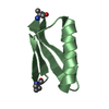
| Structure viewer | Molecule: MolmilJmol/JSmol |
|---|
- Downloads & links
-Download
| PDBx/mmCIF format | 5t7r.cif.gz | 54.9 KB | Display | PDBx/mmCIF format |
|---|---|---|---|---|
| PDB format | pdb5t7r.ent.gz | 43.6 KB | Display | PDB format |
| PDBx/mmJSON format | 5t7r.json.gz | Tree view | PDBx/mmJSON format | |
| Others |  Other downloads Other downloads |
-Validation report
| Arichive directory | https://data.pdbj.org/pub/pdb/validation_reports/t7/5t7rftp://data.pdbj.org/pub/pdb/validation_reports/t7/5t7r | HTTPS FTP |
|---|
-Related structure data
| Similar structure data |
|---|

-Links
 PDBj
PDBj


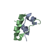
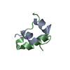
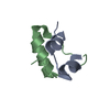
- Assembly
| Deposited unit | 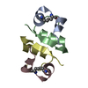
| |||||||||||||||
|---|---|---|---|---|---|---|---|---|---|---|---|---|---|---|---|---|
| 1 | 
| |||||||||||||||
| 2 | 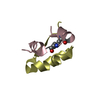
| |||||||||||||||
| Unit cell |
| |||||||||||||||
| Components on special symmetry positions |
|
-Components
| #1: Protein/peptide | Mass: 2347.622 Da / Num. of mol.: 2 / Mutation: C6(ABA), C11(ABA) / Source method: obtained synthetically Details: A chain of human insulin with CysA6 and CysA11 replaced with alpha-aminobutyric acid residues with a trans-dicarba bond formed between their respective gamma-carbons Source: (synth.) Homo sapiens (human) / References: UniProt: P01308#2: Protein/peptide | Mass: 3433.953 Da / Num. of mol.: 2 / Source method: obtained synthetically / Source: (synth.) Homo sapiens (human) / References: UniProt: P01308#3: Water | ChemComp-HOH / | Water Mass: 18.015 Da / Num. of mol.: 97 / Source method: isolated from a natural source / Formula: H2O Mass: 18.015 Da / Num. of mol.: 97 / Source method: isolated from a natural source / Formula: H2O |
|---|
-Experimental details
-Experiment
| Experiment | Method: X-RAY DIFFRACTION / Number of used crystals: 1 |
|---|
- Sample preparation
| Crystal | Density Matthews: 3.33 Å3/Da / Density % sol: 63.02 % |
|---|---|
| Crystal grow | Temperature: 300 K / Method: vapor diffusion, hanging drop / pH: 8.5 Details: 0.9 M potassium sodium tartrate, 0.1 M Tris HCl pH 8.5, 0.5% PEG 5000 MME |
-Data collection
| Diffraction | Mean temperature: 100 K |
|---|---|
| Diffraction source | Source: SYNCHROTRON / Site: Australian Synchrotron  / Beamline: MX2 / Wavelength: 0.9537 Å / Beamline: MX2 / Wavelength: 0.9537 Å |
| Detector | Type: ADSC QUANTUM 315r / Detector: CCD / Date: Sep 1, 2016 |
| Radiation | Protocol: SINGLE WAVELENGTH / Monochromatic (M) / Laue (L): M / Scattering type: x-ray |
| Radiation wavelength | Wavelength: 0.9537 Å / Relative weight: 1 |
| Reflection | Resolution: 1.55→30 Å / Num. obs: 22449 / % possible obs: 99.9 % / Redundancy: 10.8 % / CC1/2: 0.999 / Rmerge(I) obs: 0.144 / Net I/σ(I): 11.7 |
| Reflection shell | Resolution: 1.55→1.6 Å / Redundancy: 10.4 % / Rmerge(I) obs: 5.03 / Mean I/σ(I) obs: 0.38 / CC1/2: 0.11 / % possible all: 100 |
- Processing
| Software |
| |||||||||||||||||||||||||||||||||||||||||||||||||||||||||||||||
|---|---|---|---|---|---|---|---|---|---|---|---|---|---|---|---|---|---|---|---|---|---|---|---|---|---|---|---|---|---|---|---|---|---|---|---|---|---|---|---|---|---|---|---|---|---|---|---|---|---|---|---|---|---|---|---|---|---|---|---|---|---|---|---|---|
| Refinement | Resolution: 1.55→19.32 Å / SU ML: 0.26 / Cross valid method: FREE R-VALUE / σ(F): 1.33 / Phase error: 27.47 / Stereochemistry target values: ML
| |||||||||||||||||||||||||||||||||||||||||||||||||||||||||||||||
| Solvent computation | Shrinkage radii: 0.9 Å / VDW probe radii: 1.11 Å / Solvent model: FLAT BULK SOLVENT MODEL | |||||||||||||||||||||||||||||||||||||||||||||||||||||||||||||||
| Refinement step | Cycle: LAST / Resolution: 1.55→19.32 Å
| |||||||||||||||||||||||||||||||||||||||||||||||||||||||||||||||
| Refine LS restraints |
| |||||||||||||||||||||||||||||||||||||||||||||||||||||||||||||||
| LS refinement shell |
| |||||||||||||||||||||||||||||||||||||||||||||||||||||||||||||||
| Refinement TLS params. | Method: refined / Origin x: 97.0259 Å / Origin y: 38.9391 Å / Origin z: -0.7338 Å
| |||||||||||||||||||||||||||||||||||||||||||||||||||||||||||||||
| Refinement TLS group | Selection details: all |
