 Movie
Movie Controller

[English] 日本語
 Yorodumi
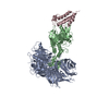
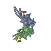
- PDB-6bn7: Crystal structure of DDB1-CRBN-BRD4(BD1) complex bound to dBET23 ... -
+ Open data

- Basic information
| Entry | Database: PDB / ID: 6bn7 | ||||||||||||||||||
|---|---|---|---|---|---|---|---|---|---|---|---|---|---|---|---|---|---|---|---|
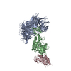
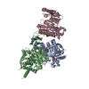
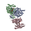
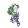
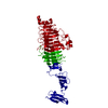
| Title | Crystal structure of DDB1-CRBN-BRD4(BD1) complex bound to dBET23 PROTAC. | ||||||||||||||||||
 Components Components |
| ||||||||||||||||||
 Keywords Keywords |  LIGASE / PROTAC / degrader / E3 ligase / CRBN LIGASE / PROTAC / degrader / E3 ligase / CRBN | ||||||||||||||||||
| Function / homology |  Function and homology information Function and homology informationnegative regulation of monoatomic ion transmembrane transport / positive regulation by virus of viral protein levels in host cell / epigenetic programming in the zygotic pronuclei / spindle assembly involved in female meiosis / Cul4-RING E3 ubiquitin ligase complex / UV-damage excision repair / biological process involved in interaction with symbiont / regulation of mitotic cell cycle phase transition / WD40-repeat domain binding / Cul4A-RING E3 ubiquitin ligase complex ...negative regulation of monoatomic ion transmembrane transport / positive regulation by virus of viral protein levels in host cell / epigenetic programming in the zygotic pronuclei / spindle assembly involved in female meiosis / Cul4-RING E3 ubiquitin ligase complex / UV-damage excision repair / biological process involved in interaction with symbiont / regulation of mitotic cell cycle phase transition / WD40-repeat domain binding / Cul4A-RING E3 ubiquitin ligase complex / Cul4B-RING E3 ubiquitin ligase complex / ubiquitin ligase complex scaffold activity / negative regulation of reproductive process / negative regulation of developmental process / locomotory exploration behavior / cullin family protein binding / RNA polymerase II C-terminal domain binding / negative regulation of DNA damage checkpoint / positive regulation of Wnt signaling pathway / P-TEFb complex binding / viral release from host cell / negative regulation by host of viral transcription / ectopic germ cell programmed cell death / positive regulation of T-helper 17 cell lineage commitment / negative regulation of protein-containing complex assembly / positive regulation of viral genome replication / positive regulation of gluconeogenesis / positive regulation of G2/M transition of mitotic cell cycle / histone reader activity / RNA polymerase II CTD heptapeptide repeat kinase activity / condensed nuclear chromosome / proteasomal protein catabolic process / nucleotide-excision repair / Recognition of DNA damage by PCNA-containing replication complex / positive regulation of protein-containing complex assembly / positive regulation of transcription elongation by RNA polymerase II / transcription coregulator activity / DNA Damage Recognition in GG-NER / lysine-acetylated histone binding / regulation of circadian rhythm / Dual Incision in GG-NER / Transcription-Coupled Nucleotide Excision Repair (TC-NER) / Formation of TC-NER Pre-Incision Complex / Wnt signaling pathway / Formation of Incision Complex in GG-NER / Dual incision in TC-NER / Gap-filling DNA repair synthesis and ligation in TC-NER / positive regulation of protein catabolic process / cellular response to UV / rhythmic process / protein-macromolecule adaptor activity / p53 binding / site of double-strand break / Neddylation / chromosome / regulation of inflammatory response / ubiquitin-dependent protein catabolic process / proteasome-mediated ubiquitin-dependent protein catabolic process / positive regulation of canonical NF-kappaB signal transduction / Potential therapeutics for SARS / transmembrane transporter binding / chromosome, telomeric region / damaged DNA binding / transcription coactivator activity / protein ubiquitination / transcription cis-regulatory region binding / chromatin remodeling / DNA repair / apoptotic process / DNA damage response / chromatin binding / protein-containing complex binding / nucleolus / regulation of transcription by RNA polymerase II / negative regulation of apoptotic process / perinuclear region of cytoplasm / positive regulation of DNA-templated transcription / enzyme binding / positive regulation of transcription by RNA polymerase II / protein-containing complex / DNA binding / extracellular space / extracellular exosome / nucleoplasm / membrane / metal ion binding / nucleus / cytosol / cytoplasmSimilarity search - Function | ||||||||||||||||||
| Biological species |  Homo sapiens (human) Homo sapiens (human) | ||||||||||||||||||
| Method | X-RAY DIFFRACTION / SYNCHROTRON / MOLECULAR REPLACEMENT / Resolution: 3.501 Å | ||||||||||||||||||
 Authors Authors | Nowak, R.P. / DeAngelo, S.L. / Buckley, D. / Bradner, J.E. / Fischer, E.S. | ||||||||||||||||||
| Funding support |  United States, 5items United States, 5items
| ||||||||||||||||||
 Citation Citation | Journal: Nat. Chem. Biol. / Year: 2018 Title: Plasticity in binding confers selectivity in ligand-induced protein degradation. Authors: Nowak, R.P. / DeAngelo, S.L. / Buckley, D. / He, Z. / Donovan, K.A. / An, J. / Safaee, N. / Jedrychowski, M.P. / Ponthier, C.M. / Ishoey, M. / Zhang, T. / Mancias, J.D. / Gray, N.S. / ...Authors: Nowak, R.P. / DeAngelo, S.L. / Buckley, D. / He, Z. / Donovan, K.A. / An, J. / Safaee, N. / Jedrychowski, M.P. / Ponthier, C.M. / Ishoey, M. / Zhang, T. / Mancias, J.D. / Gray, N.S. / Bradner, J.E. / Fischer, E.S. | ||||||||||||||||||
| History |
|
- Structure visualization
| Structure viewer | Molecule: MolmilJmol/JSmol |
|---|
- Downloads & links
-Download
| PDBx/mmCIF format | 6bn7.cif.gz | 554.4 KB | Display | PDBx/mmCIF format |
|---|---|---|---|---|
| PDB format | pdb6bn7.ent.gz | 455.2 KB | Display | PDB format |
| PDBx/mmJSON format | 6bn7.json.gz | Tree view | PDBx/mmJSON format | |
| Others |  Other downloads Other downloads |
-Validation report
| Arichive directory | https://data.pdbj.org/pub/pdb/validation_reports/bn/6bn7ftp://data.pdbj.org/pub/pdb/validation_reports/bn/6bn7 | HTTPS FTP |
|---|
-Related structure data
| Related structure data |  6bn8C  6bn9C 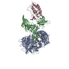 6bnbC  6boyC 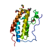 3mxfS 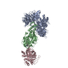 5fqdS S: Starting model for refinement C: citing same article ( |
|---|---|
| Similar structure data |

-Links
 PDBj
PDBj


- Assembly
| Deposited unit | 
| ||||||||
|---|---|---|---|---|---|---|---|---|---|
| 1 |
| ||||||||
| Unit cell |
|
-Components
| #1: Protein | Mass: 96425.586 Da / Num. of mol.: 1 Source method: isolated from a genetically manipulated source Source: (gene. exp.) Homo sapiens (human) / Gene: DDB1, XAP1 / Production host:  Trichoplusia ni (cabbage looper) / References: UniProt: Q16531 Trichoplusia ni (cabbage looper) / References: UniProt: Q16531 |
|---|---|
| #2: Protein | Mass: 53005.207 Da / Num. of mol.: 1 Source method: isolated from a genetically manipulated source Source: (gene. exp.) Homo sapiens (human) / Gene: CRBN, AD-006 / Production host: Trichoplusia ni (cabbage looper) / References: UniProt: Q96SW2 |
| #3: Protein | BRD4 / Protein HUNK1 Mass: 15099.380 Da / Num. of mol.: 1 / Mutation: T43M Source method: isolated from a genetically manipulated source Source: (gene. exp.) Homo sapiens (human) / Gene: BRD4, HUNK1 / Production host:  Escherichia coli (E. coli) / References: UniProt: O60885 Escherichia coli (E. coli) / References: UniProt: O60885 |
| #4: Chemical | ChemComp-ZN /   Mass: 65.409 Da / Num. of mol.: 1 / Source method: obtained synthetically / Formula: Zn Mass: 65.409 Da / Num. of mol.: 1 / Source method: obtained synthetically / Formula: Zn |
| #5: Chemical | ChemComp-RN3 /   Mass: 885.384 Da / Num. of mol.: 1 / Source method: obtained synthetically / Formula: C43H45ClN8O9S / Feature type: SUBJECT OF INVESTIGATION Mass: 885.384 Da / Num. of mol.: 1 / Source method: obtained synthetically / Formula: C43H45ClN8O9S / Feature type: SUBJECT OF INVESTIGATION |
-Experimental details
-Experiment
| Experiment | Method: X-RAY DIFFRACTION / Number of used crystals: 1 |
|---|
- Sample preparation
| Crystal | Density Matthews: 3.71 Å3/Da / Density % sol: 66.89 % |
|---|---|
| Crystal grow | Temperature: 293 K / Method: vapor diffusion, sitting drop / pH: 8.5 Details: 9% (w/v) PEG20K, 18% (v/v) PEG MME 550, 0.09M BICINE pH8.5, 9% Silver bullet B5 (0.33% w/v 2,7-Naphthalenedisulfonic acid disodium salt, 0.33% w/v Azelaic acid, 0.33% w/v trans-Cinnamic ...Details: 9% (w/v) PEG20K, 18% (v/v) PEG MME 550, 0.09M BICINE pH8.5, 9% Silver bullet B5 (0.33% w/v 2,7-Naphthalenedisulfonic acid disodium salt, 0.33% w/v Azelaic acid, 0.33% w/v trans-Cinnamic acid, 0.02 M HEPES sodium pH 6.8) |
-Data collection
| Diffraction | Mean temperature: 100 K |
|---|---|
| Diffraction source | Source: SYNCHROTRON / Site: APS / Beamline: 24-ID-C / Wavelength: 0.9791 Å |
| Detector | Type: DECTRIS PILATUS 6M-F / Detector: PIXEL / Date: Oct 20, 2016 |
| Radiation | Monochromator: Si(111) / Protocol: SINGLE WAVELENGTH / Monochromatic (M) / Laue (L): M / Scattering type: x-ray |
| Radiation wavelength | Wavelength: 0.9791 Å / Relative weight: 1 |
| Reflection | Resolution: 3.49→49.87 Å / Num. obs: 31460 / % possible obs: 99.5 % / Redundancy: 11.4 % / CC1/2: 0.999 / Rmerge(I) obs: 0.128 / Rpim(I) all: 0.055 / Rrim(I) all: 0.14 / Net I/σ(I): 11.4 |
| Reflection shell | Resolution: 3.49→3.68 Å / Redundancy: 10.5 % / Rmerge(I) obs: 3.561 / Mean I/σ(I) obs: 0.7 / Num. unique obs: 4335 / CC1/2: 0.328 / Rpim(I) all: 1.173 / Rrim(I) all: 3.924 / % possible all: 97.3 |
- Processing
| Software |
| |||||||||||||||||||||||||||||||||||||||||||||||||||||||||||||||||||||||||||||
|---|---|---|---|---|---|---|---|---|---|---|---|---|---|---|---|---|---|---|---|---|---|---|---|---|---|---|---|---|---|---|---|---|---|---|---|---|---|---|---|---|---|---|---|---|---|---|---|---|---|---|---|---|---|---|---|---|---|---|---|---|---|---|---|---|---|---|---|---|---|---|---|---|---|---|---|---|---|---|
| Refinement | Method to determine structure: MOLECULAR REPLACEMENT Starting model: 3mxf, 5fqd Resolution: 3.501→49.868 Å / SU ML: 0.55 / Cross valid method: FREE R-VALUE / σ(F): 1.33 / Phase error: 27.83
| |||||||||||||||||||||||||||||||||||||||||||||||||||||||||||||||||||||||||||||
| Solvent computation | Shrinkage radii: 0.9 Å / VDW probe radii: 1.11 Å | |||||||||||||||||||||||||||||||||||||||||||||||||||||||||||||||||||||||||||||
| Refinement step | Cycle: LAST / Resolution: 3.501→49.868 Å
| |||||||||||||||||||||||||||||||||||||||||||||||||||||||||||||||||||||||||||||
| Refine LS restraints |
| |||||||||||||||||||||||||||||||||||||||||||||||||||||||||||||||||||||||||||||
| LS refinement shell |
| |||||||||||||||||||||||||||||||||||||||||||||||||||||||||||||||||||||||||||||
| Refinement TLS params. | Method: refined / Origin x: 67.9938 Å / Origin y: 54.2194 Å / Origin z: 8.302 Å
| |||||||||||||||||||||||||||||||||||||||||||||||||||||||||||||||||||||||||||||
| Refinement TLS group | Selection details: all |
