- PDB-5fqd: Structural basis of Lenalidomide induced CK1a degradation by the ... -
+
Open data
ID or keywords:
Loading...
-
Basic information
Entry
Database: PDB / ID: 5fqd
Title
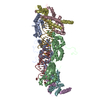
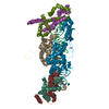
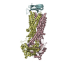
Structural basis of Lenalidomide induced CK1a degradation by the crl4crbn ubiquitin ligase
Components
CASEIN KINASE I ISOFORM ALPHA
DNA DAMAGE-BINDING PROTEIN 1
PROTEIN CEREBLON
Keywords
LIGASE / DNA BINDING
Function / homology
Function and homology information
negative regulation of monoatomic ion transmembrane transport / Activation of SMO / intermediate filament cytoskeleton organization / negative regulation of NLRP3 inflammasome complex assembly / cellular response to nutrient / positive regulation by virus of viral protein levels in host cell / APC truncation mutants have impaired AXIN binding / AXIN missense mutants destabilize the destruction complex / Truncations of AMER1 destabilize the destruction complex / epigenetic programming in the zygotic pronuclei ...negative regulation of monoatomic ion transmembrane transport / Activation of SMO / intermediate filament cytoskeleton organization / negative regulation of NLRP3 inflammasome complex assembly / cellular response to nutrient / positive regulation by virus of viral protein levels in host cell / APC truncation mutants have impaired AXIN binding / AXIN missense mutants destabilize the destruction complex / Truncations of AMER1 destabilize the destruction complex / epigenetic programming in the zygotic pronuclei / spindle assembly involved in female meiosis / beta-catenin destruction complex / Cul4-RING E3 ubiquitin ligase complex / Beta-catenin phosphorylation cascade / Signaling by GSK3beta mutants / CTNNB1 S33 mutants aren't phosphorylated / CTNNB1 S37 mutants aren't phosphorylated / CTNNB1 S45 mutants aren't phosphorylated / CTNNB1 T41 mutants aren't phosphorylated / UV-damage excision repair / Disassembly of the destruction complex and recruitment of AXIN to the membrane / biological process involved in interaction with symbiont / Maturation of nucleoprotein / regulation of mitotic cell cycle phase transition / WD40-repeat domain binding / Cul4A-RING E3 ubiquitin ligase complex / Cul4B-RING E3 ubiquitin ligase complex / ubiquitin ligase complex scaffold activity / positive regulation of Rho protein signal transduction / negative regulation of reproductive process / negative regulation of developmental process / Golgi organization / locomotory exploration behavior / cullin family protein binding / positive regulation of Wnt signaling pathway / viral release from host cell / ectopic germ cell programmed cell death / negative regulation of protein-containing complex assembly / positive regulation of viral genome replication / positive regulation of gluconeogenesis / positive regulation of TORC1 signaling / ciliary basal body / proteasomal protein catabolic process / nucleotide-excision repair / Recognition of DNA damage by PCNA-containing replication complex / positive regulation of protein-containing complex assembly / DNA Damage Recognition in GG-NER / Degradation of GLI2 by the proteasome / GLI3 is processed to GLI3R by the proteasome / Degradation of beta-catenin by the destruction complex / negative regulation of canonical Wnt signaling pathway / cilium / regulation of circadian rhythm / Dual Incision in GG-NER / Transcription-Coupled Nucleotide Excision Repair (TC-NER) / kinetochore / spindle / Formation of TC-NER Pre-Incision Complex / Wnt signaling pathway / Formation of Incision Complex in GG-NER / Dual incision in TC-NER / Gap-filling DNA repair synthesis and ligation in TC-NER / positive regulation of protein catabolic process / cellular response to UV / rhythmic process / protein-macromolecule adaptor activity / positive regulation of proteasomal ubiquitin-dependent protein catabolic process / site of double-strand break / Neddylation / ubiquitin-dependent protein catabolic process / proteasome-mediated ubiquitin-dependent protein catabolic process / peptidyl-serine phosphorylation / Potential therapeutics for SARS / transmembrane transporter binding / chromosome, telomeric region / damaged DNA binding / cell surface receptor signaling pathway / protein ubiquitination / viral protein processing / non-specific serine/threonine protein kinase / protein kinase activity / nuclear speck / cell cycle / cell division / protein phosphorylation / protein serine kinase activity / DNA repair / protein serine/threonine kinase activity / centrosome / apoptotic process / DNA damage response / protein-containing complex binding / nucleolus / negative regulation of apoptotic process / perinuclear region of cytoplasm / signal transduction / protein-containing complex / DNA binding / extracellular space / extracellular exosome Similarity search - Function
Methane Monooxygenase Hydroxylase; Chain G, domain 1 - #1480 / LON domain-like / Peptide methionine sulfoxide reductase. / DNA polymerase; domain 1 - #910 / Yippee/Mis18/Cereblon / Yippee zinc-binding/DNA-binding /Mis18, centromere assembly / Metal Binding Protein, Guanine Nucleotide Exchange Factor; Chain A / Archaeosine Trna-guanine Transglycosylase; Chain: A, domain 4 / CULT domain / CULT domain profile. ...Methane Monooxygenase Hydroxylase; Chain G, domain 1 - #1480 / LON domain-like / Peptide methionine sulfoxide reductase. / DNA polymerase; domain 1 - #910 / Yippee/Mis18/Cereblon / Yippee zinc-binding/DNA-binding /Mis18, centromere assembly / Metal Binding Protein, Guanine Nucleotide Exchange Factor; Chain A / Archaeosine Trna-guanine Transglycosylase; Chain: A, domain 4 / CULT domain / CULT domain profile. / Lon protease, N-terminal domain superfamily / Lon N-terminal domain profile. / Lon protease, N-terminal domain / ATP-dependent protease La (LON) substrate-binding domain / Found in ATP-dependent protease La (LON) / Cleavage/polyadenylation specificity factor, A subunit, N-terminal / Mono-functional DNA-alkylating methyl methanesulfonate N-term / Cleavage/polyadenylation specificity factor, A subunit, C-terminal / CPSF A subunit region / PUA-like superfamily / YVTN repeat-like/Quinoprotein amine dehydrogenase / 7 Propeller / Methylamine Dehydrogenase; Chain H / Methane Monooxygenase Hydroxylase; Chain G, domain 1 / Beta Complex / DNA polymerase; domain 1 / Roll / WD40-repeat-containing domain superfamily / Transferase(Phosphotransferase) domain 1 / Transferase(Phosphotransferase); domain 1 / Phosphorylase Kinase; domain 1 / Phosphorylase Kinase; domain 1 / Serine/threonine-protein kinase, active site / Serine/Threonine protein kinases active-site signature. / WD40/YVTN repeat-like-containing domain superfamily / Protein kinase domain / Serine/Threonine protein kinases, catalytic domain / Protein kinase, ATP binding site / Protein kinases ATP-binding region signature. / Protein kinase domain profile. / Protein kinase domain / Protein kinase-like domain superfamily / Up-down Bundle / 2-Layer Sandwich / Orthogonal Bundle / Mainly Beta / Mainly Alpha / Alpha Beta Similarity search - Domain/homology
S-Lenalidomide / Casein kinase I isoform alpha / DNA damage-binding protein 1 / Protein cereblon Similarity search - Component
A: DNA DAMAGE-BINDING PROTEIN 1 B: PROTEIN CEREBLON C: CASEIN KINASE I ISOFORM ALPHA D: DNA DAMAGE-BINDING PROTEIN 1 E: PROTEIN CEREBLON F: CASEIN KINASE I ISOFORM ALPHA hetero molecules
DNADAMAGE-BINDINGPROTEIN1 / DDB P127 SUBUNIT / DNA DAMAGE-BINDING PROTEIN A / DDBA / DAMAGE -SPECIFIC DNA-BINDING PROTEIN 1 / ...DDB P127 SUBUNIT / DNA DAMAGE-BINDING PROTEIN A / DDBA / DAMAGE -SPECIFIC DNA-BINDING PROTEIN 1 / HBV X-ASSOCIATED PROTEIN 1 / XAP-1 / UV-DAMAGED DNA-BINDING FACTOR / UV-DAMAGED DNA-BINDING PROTEIN 1 / UV-DDB 1 / XPE-BINDING FACTOR / XPE-BF / XERODERMA PIGMENTOSUM GROUP E-COMPLEMENTING PROTEIN / XPCE
Mass: 95773.695 Da / Num. of mol.: 2 Source method: isolated from a genetically manipulated source Source: (gene. exp.) HOMO SAPIENS (human) / Cell line (production host): High Five / Production host: TRICHOPLUSIA NI (cabbage looper) / References: UniProt: Q16531, ubiquitin-protein ligase
#2: Protein
PROTEINCEREBLON
Mass: 48976.117 Da / Num. of mol.: 2 Source method: isolated from a genetically manipulated source Source: (gene. exp.) HOMO SAPIENS (human) / Cell line (production host): High Five / Production host: TRICHOPLUSIA NI (cabbage looper) / References: UniProt: Q96SW2
Mass: 39302.238 Da / Num. of mol.: 2 Source method: isolated from a genetically manipulated source Source: (gene. exp.) HOMO SAPIENS (human) / Cell line (production host): High Five / Production host: TRICHOPLUSIA NI (cabbage looper) / References: UniProt: P48729
In the structure databanks used in Yorodumi, some data are registered as the other names, "COVID-19 virus" and "2019-nCoV". Here are the details of the virus and the list of structure data.
Jan 31, 2019. EMDB accession codes are about to change! (news from PDBe EMDB page)
EMDB accession codes are about to change! (news from PDBe EMDB page)
The allocation of 4 digits for EMDB accession codes will soon come to an end. Whilst these codes will remain in use, new EMDB accession codes will include an additional digit and will expand incrementally as the available range of codes is exhausted. The current 4-digit format prefixed with “EMD-” (i.e. EMD-XXXX) will advance to a 5-digit format (i.e. EMD-XXXXX), and so on. It is currently estimated that the 4-digit codes will be depleted around Spring 2019, at which point the 5-digit format will come into force.
The EM Navigator/Yorodumi systems omit the EMD- prefix.
Related info.:Q: What is EMD? / ID/Accession-code notation in Yorodumi/EM Navigator
Yorodumi is a browser for structure data from EMDB, PDB, SASBDB, etc.
This page is also the successor to EM Navigator detail page, and also detail information page/front-end page for Omokage search.
The word "yorodu" (or yorozu) is an old Japanese word meaning "ten thousand". "mi" (miru) is to see.
Related info.:EMDB / PDB / SASBDB / Comparison of 3 databanks / Yorodumi Search / Aug 31, 2016. New EM Navigator & Yorodumi / Yorodumi Papers / Jmol/JSmol / Function and homology information / Changes in new EM Navigator and Yorodumi
 Movie
Movie Controller

 Yorodumi
 Open data
 Basic information
 Components
 Keywords
 LIGASE /
  Function and homology information
 Authors
 Citation
 Structure visualization
 Downloads & links
 Other downloads
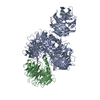


 PDBj
PDBj


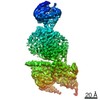
 Assembly


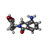

 Mass: 65.409 Da / Num. of mol.: 2 / Source method: obtained synthetically / Formula: Zn
 Mass: 259.261 Da / Num. of mol.: 2 / Source method: obtained synthetically / Formula: C13H13N3O3
 Sample preparation
 / Beamline: X10SA / Wavelength: 1.28161
  Processing