 Movie
Movie Controller


+ Open data

- Basic information
| Entry | Database: PDB / ID: 6bk9 | ||||||
|---|---|---|---|---|---|---|---|
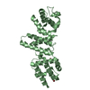
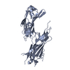
| Title | Crystal Structure of Squid Arrestin | ||||||
 Components Components | Visual arrestin | ||||||
 Keywords Keywords |  SIGNALING PROTEIN / Arrestin / Phosphorylation independent / Squid / invertebrate / Rhodopsin / adapter protein SIGNALING PROTEIN / Arrestin / Phosphorylation independent / Squid / invertebrate / Rhodopsin / adapter protein | ||||||
| Function / homology |  Function and homology information Function and homology information | ||||||
| Biological species |  Doryteuthis pealeii (longfin inshore squid) Doryteuthis pealeii (longfin inshore squid) | ||||||
| Method | X-RAY DIFFRACTION / SYNCHROTRON / MOLECULAR REPLACEMENT / Resolution: 3.00005573983 Å | ||||||
 Authors Authors | Eger, B.T. / Bandyopadhyay, A. / Yedidi, R.S. / Ernst, O.P. | ||||||
| Funding support |  Canada, 1items Canada, 1items
| ||||||
 Citation Citation | Journal: J. Mol. Biol. / Year: 2018 Title: A Novel Polar Core and Weakly Fixed C-Tail in Squid Arrestin Provide New Insight into Interaction with Rhodopsin. Authors: Bandyopadhyay, A. / Van Eps, N. / Eger, B.T. / Rauscher, S. / Yedidi, R.S. / Moroni, T. / West, G.M. / Robinson, K.A. / Griffin, P.R. / Mitchell, J. / Ernst, O.P. | ||||||
| History |
|
- Structure visualization
| Structure viewer | Molecule: MolmilJmol/JSmol |
|---|
- Downloads & links
-Download
| PDBx/mmCIF format | 6bk9.cif.gz | 143.8 KB | Display | PDBx/mmCIF format |
|---|---|---|---|---|
| PDB format | pdb6bk9.ent.gz | 92.9 KB | Display | PDB format |
| PDBx/mmJSON format | 6bk9.json.gz | Tree view | PDBx/mmJSON format | |
| Others |  Other downloads Other downloads |
-Validation report
| Arichive directory | https://data.pdbj.org/pub/pdb/validation_reports/bk/6bk9ftp://data.pdbj.org/pub/pdb/validation_reports/bk/6bk9 | HTTPS FTP |
|---|
-Related structure data
| Related structure data | 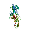 1jsyS S: Starting model for refinement |
|---|---|
| Similar structure data |


-Links
 PDBj
PDBj
- Assembly
| Deposited unit | 
| ||||||||||||
|---|---|---|---|---|---|---|---|---|---|---|---|---|---|
| 1 |
| ||||||||||||
| Unit cell |
|
-Components
| #1: Protein | Mass: 42974.117 Da / Num. of mol.: 1 Source method: isolated from a genetically manipulated source Source: (gene. exp.) Doryteuthis pealeii (longfin inshore squid)Production host:  Escherichia coli (E. coli) / References: UniProt: Q963B5 Escherichia coli (E. coli) / References: UniProt: Q963B5 |
|---|---|
| #2: Chemical | ChemComp-CL / Chloride  Mass: 35.453 Da / Num. of mol.: 1 / Source method: obtained synthetically / Formula: Cl Mass: 35.453 Da / Num. of mol.: 1 / Source method: obtained synthetically / Formula: Cl |
-Experimental details
-Experiment
| Experiment | Method: X-RAY DIFFRACTION / Number of used crystals: 1 |
|---|
- Sample preparation
| Crystal | Density Matthews: 2.76 Å3/Da / Density % sol: 55.5 % |
|---|---|
| Crystal grow | Temperature: 293 K / Method: vapor diffusion, sitting drop / pH: 6.5 Details: 1.2 M Ammonium Sulphate, 300 mM KH2PO4, 100 mM Bis-Tris pH 6.5 |
-Data collection
| Diffraction | Mean temperature: 100 K |
|---|---|
| Diffraction source | Source: SYNCHROTRON / Site: APS  / Beamline: 17-ID / Wavelength: 1 Å / Beamline: 17-ID / Wavelength: 1 Å |
| Detector | Type: DECTRIS PILATUS3 6M / Detector: PIXEL / Date: Aug 25, 2015 |
| Radiation | Protocol: SINGLE WAVELENGTH / Monochromatic (M) / Laue (L): M / Scattering type: x-ray |
| Radiation wavelength | Wavelength: 1 Å / Relative weight: 1 |
| Reflection | Resolution: 2.88→31 Å / Num. obs: 11540 / % possible obs: 99.5 % / Redundancy: 10.8 % / Biso Wilson estimate: 92.8591770063 Å2 / CC1/2: 0.995 / Net I/σ(I): 13.9 |
- Processing
| Software |
| ||||||||||||||||||||||||||||||||||||||||
|---|---|---|---|---|---|---|---|---|---|---|---|---|---|---|---|---|---|---|---|---|---|---|---|---|---|---|---|---|---|---|---|---|---|---|---|---|---|---|---|---|---|
| Refinement | Method to determine structure: MOLECULAR REPLACEMENT Starting model: 1JSY Resolution: 3.00005573983→31.0041375064 Å / SU ML: 0.499771948767 / Cross valid method: THROUGHOUT / σ(F): 1.35576831605 / Phase error: 41.0895765951
| ||||||||||||||||||||||||||||||||||||||||
| Solvent computation | Shrinkage radii: 0.9 Å / VDW probe radii: 1.11 Å | ||||||||||||||||||||||||||||||||||||||||
| Displacement parameters | Biso mean: 90.8163449963 Å2 | ||||||||||||||||||||||||||||||||||||||||
| Refinement step | Cycle: LAST / Resolution: 3.00005573983→31.0041375064 Å
| ||||||||||||||||||||||||||||||||||||||||
| Refine LS restraints |
| ||||||||||||||||||||||||||||||||||||||||
| LS refinement shell |
| ||||||||||||||||||||||||||||||||||||||||
| Refinement TLS params. | Method: refined / Origin x: 28.0778725325 Å / Origin y: -19.5949687097 Å / Origin z: -10.2539815501 Å
| ||||||||||||||||||||||||||||||||||||||||
| Refinement TLS group | Selection details: all |
