 Movie
Movie Controller

[English] 日本語
 Yorodumi
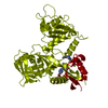
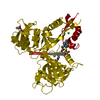
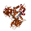
- PDB-6b0u: Crystal structure of acetyltransferase Eis from Mycobacterium tub... -
+ Open data

- Basic information
| Entry | Database: PDB / ID: 6b0u | ||||||
|---|---|---|---|---|---|---|---|
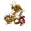
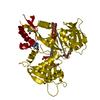
| Title | Crystal structure of acetyltransferase Eis from Mycobacterium tuberculosis in complex with a Lys-containing peptide | ||||||
 Components Components |
| ||||||
 Keywords Keywords |  TRANSFERASE / histone acetylation / aminoglycoside / drug resistance / tuberculosis / acetylation TRANSFERASE / histone acetylation / aminoglycoside / drug resistance / tuberculosis / acetylation | ||||||
| Function / homology |  Function and homology information Function and homology informationeffector-mediated defense to host-produced reactive oxygen species / symbiont-mediated perturbation of host programmed cell death / symbiont-mediated perturbation of host inflammatory response / symbiont-mediated suppression of host defense-related programmed cell death / aminoglycoside antibiotic catabolic process / aminoglycoside N-acetyltransferase activity / symbiont-mediated perturbation of host innate immune response / Suppression of autophagy / bacterial extracellular vesicle / biological process involved in interaction with host ...effector-mediated defense to host-produced reactive oxygen species / symbiont-mediated perturbation of host programmed cell death / symbiont-mediated perturbation of host inflammatory response / symbiont-mediated suppression of host defense-related programmed cell death / aminoglycoside antibiotic catabolic process / aminoglycoside N-acetyltransferase activity / symbiont-mediated perturbation of host innate immune response / Suppression of autophagy / bacterial extracellular vesicle / biological process involved in interaction with host / host cell cytoplasmic vesicle / N-acetyltransferase activity / peptide-lysine-N-acetyltransferase activity / Transferases; Acyltransferases; Transferring groups other than aminoacyl groups / host extracellular space / response to antibiotic / identical protein binding / cytosolSimilarity search - Function | ||||||
| Biological species |   Mycobacterium tuberculosis (bacteria)Mycobacterium tuberculosis H37Rv (bacteria) Mycobacterium tuberculosis (bacteria)Mycobacterium tuberculosis H37Rv (bacteria) | ||||||
| Method | X-RAY DIFFRACTION / SYNCHROTRON / MOLECULAR REPLACEMENT / Resolution: 2.8 Å | ||||||
 Authors Authors | Biswas, T. / Pang, A.H. / Garneau-Tsodikova, S. / Tsodikov, O.V. | ||||||
| Funding support |  United States, 1items United States, 1items
| ||||||
 Citation Citation | Journal: Biochemistry / Year: 2018 Title: Acetylation by Eis and Deacetylation by Rv1151c of Mycobacterium tuberculosis HupB: Biochemical and Structural Insight. Authors: Green, K.D. / Biswas, T. / Pang, A.H. / Willby, M.J. / Reed, M.S. / Stuchlik, O. / Pohl, J. / Posey, J.E. / Tsodikov, O.V. / Garneau-Tsodikova, S. | ||||||
| History |
|
- Structure visualization
| Structure viewer | Molecule: MolmilJmol/JSmol |
|---|
- Downloads & links
-Download
| PDBx/mmCIF format | 6b0u.cif.gz | 238.8 KB | Display | PDBx/mmCIF format |
|---|---|---|---|---|
| PDB format | pdb6b0u.ent.gz | 190.9 KB | Display | PDB format |
| PDBx/mmJSON format | 6b0u.json.gz | Tree view | PDBx/mmJSON format | |
| Others |  Other downloads Other downloads |
-Validation report
| Arichive directory | https://data.pdbj.org/pub/pdb/validation_reports/b0/6b0uftp://data.pdbj.org/pub/pdb/validation_reports/b0/6b0u | HTTPS FTP |
|---|
-Related structure data
| Related structure data |  3r1kS S: Starting model for refinement |
|---|---|
| Similar structure data |


-Links
 PDBj
PDBj


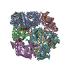
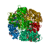
- Assembly
| Deposited unit | 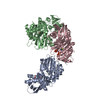
| ||||||||
|---|---|---|---|---|---|---|---|---|---|
| 1 | 
| ||||||||
| Unit cell |
|
-Components
| #1: Protein | Mass: 46692.902 Da / Num. of mol.: 3 Source method: isolated from a genetically manipulated source Source: (gene. exp.) Mycobacterium tuberculosis (strain ATCC 25618 / H37Rv) (bacteria)Strain: ATCC 25618 / H37Rv / Gene: eis, Rv2416c, MTCY253.04 / Production host: Escherichia coli BL21(DE3) (bacteria)References: UniProt: P9WFK7, Transferases; Acyltransferases; Transferring groups other than aminoacyl groups#2: Protein/peptide | Mass: 888.084 Da / Num. of mol.: 2 / Source method: obtained synthetically Source: (synth.) Mycobacterium tuberculosis H37Rv (bacteria)#3: Chemical | ChemComp-COA / Coenzyme A  Mass: 767.534 Da / Num. of mol.: 5 / Source method: obtained synthetically / Formula: C21H36N7O16P3S Mass: 767.534 Da / Num. of mol.: 5 / Source method: obtained synthetically / Formula: C21H36N7O16P3S |
|---|
-Experimental details
-Experiment
| Experiment | Method: X-RAY DIFFRACTION / Number of used crystals: 1 |
|---|
- Sample preparation
| Crystal | Density Matthews: 2.5 Å3/Da / Density % sol: 50.78 % |
|---|---|
| Crystal grow | Temperature: 295 K / Method: vapor diffusion, hanging drop / pH: 8.5 Details: peptide (1 mM) and CoA (1 mM) with protein in a 1:1 ration with reservoir solution (Tris-HCl (100 mM, pH 8.5) and PEG 8,000 (13% w/v)) |
-Data collection
| Diffraction | Mean temperature: 100 K |
|---|---|
| Diffraction source | Source: SYNCHROTRON / Site: APS / Beamline: 21-ID-G / Wavelength: 0.97 Å |
| Detector | Type: MARMOSAIC 300 mm CCD / Detector: CCD / Date: Oct 4, 2012 |
| Radiation | Protocol: SINGLE WAVELENGTH / Monochromatic (M) / Laue (L): M / Scattering type: x-ray |
| Radiation wavelength | Wavelength: 0.97 Å / Relative weight: 1 |
| Reflection | Resolution: 2.8→37.71 Å / Num. obs: 30053 / % possible obs: 87 % / Observed criterion σ(I): 2.8 / Redundancy: 2.9 % / Rmerge(I) obs: 0.06 / Net I/σ(I): 14 |
| Reflection shell | Resolution: 2.8→2.85 Å / Rmerge(I) obs: 0.24 / % possible all: 87 |
- Processing
| Software |
| ||||||||||||||||||||||||||||||||||||||||||||||||||||||||||||||||||||||||||||||||||||||||||||||||||||||||||||||||||||||||||||||||||||||||||||||||||||||||||||||||||||||||||||||||||||||
|---|---|---|---|---|---|---|---|---|---|---|---|---|---|---|---|---|---|---|---|---|---|---|---|---|---|---|---|---|---|---|---|---|---|---|---|---|---|---|---|---|---|---|---|---|---|---|---|---|---|---|---|---|---|---|---|---|---|---|---|---|---|---|---|---|---|---|---|---|---|---|---|---|---|---|---|---|---|---|---|---|---|---|---|---|---|---|---|---|---|---|---|---|---|---|---|---|---|---|---|---|---|---|---|---|---|---|---|---|---|---|---|---|---|---|---|---|---|---|---|---|---|---|---|---|---|---|---|---|---|---|---|---|---|---|---|---|---|---|---|---|---|---|---|---|---|---|---|---|---|---|---|---|---|---|---|---|---|---|---|---|---|---|---|---|---|---|---|---|---|---|---|---|---|---|---|---|---|---|---|---|---|---|---|
| Refinement | Method to determine structure: MOLECULAR REPLACEMENT Starting model: 3R1K Resolution: 2.8→37 Å / Cor.coef. Fo:Fc: 0.934 / Cor.coef. Fo:Fc free: 0.891 / SU B: 20.379 / SU ML: 0.364 / Cross valid method: THROUGHOUT / ESU R Free: 0.442 / Stereochemistry target values: MAXIMUM LIKELIHOOD / Details: HYDROGENS HAVE BEEN ADDED IN THE RIDING POSITIONS
| ||||||||||||||||||||||||||||||||||||||||||||||||||||||||||||||||||||||||||||||||||||||||||||||||||||||||||||||||||||||||||||||||||||||||||||||||||||||||||||||||||||||||||||||||||||||
| Solvent computation | Ion probe radii: 0.8 Å / Shrinkage radii: 0.8 Å / VDW probe radii: 1.2 Å / Solvent model: MASK | ||||||||||||||||||||||||||||||||||||||||||||||||||||||||||||||||||||||||||||||||||||||||||||||||||||||||||||||||||||||||||||||||||||||||||||||||||||||||||||||||||||||||||||||||||||||
| Displacement parameters | Biso mean: 59.785 Å2
| ||||||||||||||||||||||||||||||||||||||||||||||||||||||||||||||||||||||||||||||||||||||||||||||||||||||||||||||||||||||||||||||||||||||||||||||||||||||||||||||||||||||||||||||||||||||
| Refinement step | Cycle: 1 / Resolution: 2.8→37 Å
| ||||||||||||||||||||||||||||||||||||||||||||||||||||||||||||||||||||||||||||||||||||||||||||||||||||||||||||||||||||||||||||||||||||||||||||||||||||||||||||||||||||||||||||||||||||||
| Refine LS restraints |
|