 Movie
Movie Controller


+ Open data

- Basic information
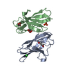
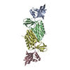
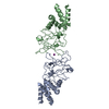
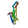
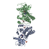
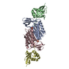
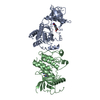
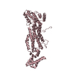
| Entry | Database: PDB / ID: 6aw3 | ||||||
|---|---|---|---|---|---|---|---|
| Title | Crystal structure of the HopQ-CEACAM3 L44Q complex | ||||||
 Components Components |
| ||||||
 Keywords Keywords |  CELL ADHESION CELL ADHESION | ||||||
| Function / homology |  Function and homology informationregulation of immune system process / specific granule membrane / protein tyrosine kinase binding / Cell surface interactions at the vascular wall / Neutrophil degranulation / cell surface / signal transduction / plasma membrane Function and homology informationregulation of immune system process / specific granule membrane / protein tyrosine kinase binding / Cell surface interactions at the vascular wall / Neutrophil degranulation / cell surface / signal transduction / plasma membraneSimilarity search - Function | ||||||
| Biological species |   Helicobacter pylori (bacteria) Helicobacter pylori (bacteria) Homo sapiens (human) Homo sapiens (human) | ||||||
| Method | X-RAY DIFFRACTION / SYNCHROTRON / MOLECULAR REPLACEMENT / Resolution: 2.66 Å | ||||||
 Authors Authors | Bonsor, D.A. / Sundberg, E.J. | ||||||
 Citation Citation | Journal: EMBO J. / Year: 2018 Title: TheHelicobacter pyloriadhesin protein HopQ exploits the dimer interface of human CEACAMs to facilitate translocation of the oncoprotein CagA. Authors: Bonsor, D.A. / Zhao, Q. / Schmidinger, B. / Weiss, E. / Wang, J. / Deredge, D. / Beadenkopf, R. / Dow, B. / Fischer, W. / Beckett, D. / Wintrode, P.L. / Haas, R. / Sundberg, E.J. | ||||||
| History |
|
- Structure visualization
| Structure viewer | Molecule: MolmilJmol/JSmol |
|---|
- Downloads & links
-Download
| PDBx/mmCIF format | 6aw3.cif.gz | 177.3 KB | Display | PDBx/mmCIF format |
|---|---|---|---|---|
| PDB format | pdb6aw3.ent.gz | 145.1 KB | Display | PDB format |
| PDBx/mmJSON format | 6aw3.json.gz | Tree view | PDBx/mmJSON format | |
| Others |  Other downloads Other downloads |
-Validation report
| Arichive directory | https://data.pdbj.org/pub/pdb/validation_reports/aw/6aw3ftp://data.pdbj.org/pub/pdb/validation_reports/aw/6aw3 | HTTPS FTP |
|---|
-Related structure data


-Links
 PDBj
PDBj- Assembly
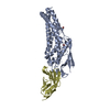
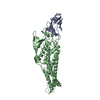
| Deposited unit | 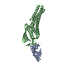
| ||||||||
|---|---|---|---|---|---|---|---|---|---|
| 1 |
| ||||||||
| 2 | 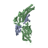
| ||||||||
| Unit cell |
|
-Components
| #1: Protein | Mass: 47027.602 Da / Num. of mol.: 1 Source method: isolated from a genetically manipulated source Source: (gene. exp.) Helicobacter pylori (bacteria) / Gene: hopQ / Production host: Escherichia coli BL21(DE3) (bacteria) / References: UniProt: H6A3H4 |
|---|---|
| #2: Protein | Mass: 12060.540 Da / Num. of mol.: 1 / Mutation: L44Q Source method: isolated from a genetically manipulated source Source: (gene. exp.) Homo sapiens (human) / Gene: CEACAM3, CD66D, CGM1 / Production host: Escherichia coli BL21(DE3) (bacteria) / Strain (production host): BL21(DE3) / References: UniProt: P40198 |
| #3: Water | ChemComp-HOH / Water Mass: 18.015 Da / Num. of mol.: 8 / Source method: isolated from a natural source / Formula: H2O Mass: 18.015 Da / Num. of mol.: 8 / Source method: isolated from a natural source / Formula: H2O |
-Experimental details
-Experiment
| Experiment | Method: X-RAY DIFFRACTION / Number of used crystals: 1 |
|---|
- Sample preparation
| Crystal | Density Matthews: 1.9 Å3/Da / Density % sol: 35.22 % |
|---|---|
| Crystal grow | Temperature: 298 K / Method: vapor diffusion, hanging drop Details: 20% w/v PEG 3000, 0.1 M Bis-Tris-HCl, pH 6.5, 0.2 M calcium acetate, 1 % v/v glycerol |
-Data collection
| Diffraction | Mean temperature: 100 K |
|---|---|
| Diffraction source | Source: SYNCHROTRON / Site: SSRL  / Beamline: BL9-2 / Wavelength: 0.97946 Å / Beamline: BL9-2 / Wavelength: 0.97946 Å |
| Detector | Type: DECTRIS PILATUS 6M / Detector: PIXEL / Date: Feb 17, 2017 Details: Rh coated flat bent M0, toroidal focusing post-monochromator M1 |
| Radiation | Monochromator: Si (111) / Protocol: SINGLE WAVELENGTH / Monochromatic (M) / Laue (L): M / Scattering type: x-ray |
| Radiation wavelength | Wavelength: 0.97946 Å / Relative weight: 1 |
| Reflection | Resolution: 2.66→102.59 Å / Num. obs: 13470 / % possible obs: 99.3 % / Redundancy: 5.6 % / Rmerge(I) obs: 0.12 / Rpim(I) all: 0.081 / Net I/σ(I): 10.7 |
| Reflection shell | Resolution: 2.66→2.79 Å / Redundancy: 5.1 % / Rmerge(I) obs: 1.179 / Mean I/σ(I) obs: 1.3 / Num. unique obs: 1699 / Rpim(I) all: 0.841 / % possible all: 96.3 |
- Processing
| Software |
| ||||||||||||||||||||||||||||||||||||||||||||||||||||||||||||||||||||||||||||||||||||||||||||||||||||||||||||||||||||||||||||||||||||||||||||||||||||||||||||||||||||||||||||||||||||||
|---|---|---|---|---|---|---|---|---|---|---|---|---|---|---|---|---|---|---|---|---|---|---|---|---|---|---|---|---|---|---|---|---|---|---|---|---|---|---|---|---|---|---|---|---|---|---|---|---|---|---|---|---|---|---|---|---|---|---|---|---|---|---|---|---|---|---|---|---|---|---|---|---|---|---|---|---|---|---|---|---|---|---|---|---|---|---|---|---|---|---|---|---|---|---|---|---|---|---|---|---|---|---|---|---|---|---|---|---|---|---|---|---|---|---|---|---|---|---|---|---|---|---|---|---|---|---|---|---|---|---|---|---|---|---|---|---|---|---|---|---|---|---|---|---|---|---|---|---|---|---|---|---|---|---|---|---|---|---|---|---|---|---|---|---|---|---|---|---|---|---|---|---|---|---|---|---|---|---|---|---|---|---|---|
| Refinement | Method to determine structure: MOLECULAR REPLACEMENT / Resolution: 2.66→102.59 Å / Cor.coef. Fo:Fc: 0.926 / Cor.coef. Fo:Fc free: 0.88 / SU B: 44.22 / SU ML: 0.393 / Cross valid method: THROUGHOUT / ESU R Free: 0.414 / Details: HYDROGENS HAVE BEEN ADDED IN THE RIDING POSITIONS
| ||||||||||||||||||||||||||||||||||||||||||||||||||||||||||||||||||||||||||||||||||||||||||||||||||||||||||||||||||||||||||||||||||||||||||||||||||||||||||||||||||||||||||||||||||||||
| Solvent computation | Ion probe radii: 0.8 Å / Shrinkage radii: 0.8 Å / VDW probe radii: 1.2 Å | ||||||||||||||||||||||||||||||||||||||||||||||||||||||||||||||||||||||||||||||||||||||||||||||||||||||||||||||||||||||||||||||||||||||||||||||||||||||||||||||||||||||||||||||||||||||
| Displacement parameters | Biso mean: 53.141 Å2
| ||||||||||||||||||||||||||||||||||||||||||||||||||||||||||||||||||||||||||||||||||||||||||||||||||||||||||||||||||||||||||||||||||||||||||||||||||||||||||||||||||||||||||||||||||||||
| Refinement step | Cycle: 1 / Resolution: 2.66→102.59 Å
| ||||||||||||||||||||||||||||||||||||||||||||||||||||||||||||||||||||||||||||||||||||||||||||||||||||||||||||||||||||||||||||||||||||||||||||||||||||||||||||||||||||||||||||||||||||||
| Refine LS restraints |
|